
Inquiry Basket
 Loading ......
Macrophages, as key components of the immune system, play a crucial role in various biological processes, including host defense, tissue repair, and immune regulation. Understanding the phenotypic characteristics and functional diversity of macrophages is essential for advancing research in immunology, cancer biology, and inflammatory diseases.
Macrophages are phagocytic immune cells that originate from monocytes and exist in various tissues throughout the body. These cells exhibit remarkable plasticity and multifunctionality, enabling them to adapt their phenotype and function according to the microenvironment they encounter. Macrophages perform a wide range of vital functions, including clearance of senescent or apoptotic cells and phagocytosis of immune complexes and pathogens. In addition to initiating immune and inflammatory responses to pathogens, macrophages also maintain tissue homeostasis and play a role in tissue repair and remodeling. Multiple studies have reported that this function is associated with many diseases, including metabolic and autoimmune diseases, cancer, infection, obesity, and fibrosis. Thus, macrophages also appear to play key roles in the tumor microenvironment, especially in matrix remodeling, angiogenesis, metastasis, and tumor progression.
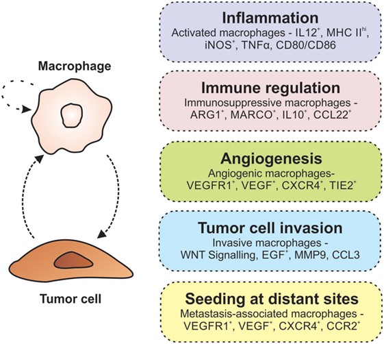 Fig. 1 Macrophages promote tumorigenesis. (Poh A R, et al., 2018)
Different factors including signaling molecules, transcription factors, growth factors, epigenetic and post-transcriptional mechanisms and changes, and niche signals such as cytokines, intercellular contacts, and metabolites lead to distinct phenotypes and macrophage activation states. In addition, macrophages can regulate their own activation status in the face of microorganisms and their products, such as lipopolysaccharides (LPS). Activation of macrophages plays a crucial role in tissue homeostasis as well as in inflammation and disease progression. In general, macrophages can be divided into two subtypes based on functions and activation state: classically activated M1 macrophages and alternatively activated M2 macrophages.
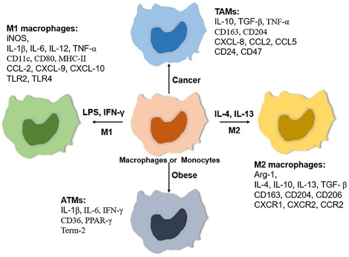 Fig. 2 Relative expressing gene profiles. (Zhang C, et al., 2021)
In the process of tissue inflammation, the M1 type appears early and recruits many inflammatory cells to fight pathogens, which is a pro-inflammatory effect. After the pathogen is eradicated, the M2 type acts to inhibit the recruitment of inflammatory cells, which is an anti-inflammatory effect. The M2 type promotes both angiogenesis and tissue repair, allowing the tissue to return to its original shape. Continuous M1 activation can cause tissue damage, leading to inflammatory diseases; M2 overactivation may lead to fibrosis due to excessive tissue repair, and even promote tumor growth through immunosuppression.
M1 macrophages are usually activated by pathogens, granulocyte-macrophage colony-stimulating factor (GM-CSF), LPS, tumor necrosis factor-alpha (TNF-α), and type 1 helper T (Th1) cytokine interferon-gamma (IFN-γ). Characteristically speaking, M1 macrophages have strong antigen-presenting activity and secrete many pro-inflammatory cytokines, such as interleukin-1 (IL-1), IL-6, TNF-α, nitric oxide (NO), and reactive oxygen species (ROS). In addition, the M1 macrophage phenotype expresses high levels of major histocompatibility complex class II (MHC II), CD68, CD80, and CD86, as well as chemokines targeting Th1 cells, including CXCL9 and CXCL12.
M2 macrophages play a role in pathogen clearance, anti-inflammatory response, and metabolism, as well as in wound healing, tissue remodeling, immune regulation, and tumor progression. These cells can be activated by parasitic or fungal infections, apoptotic cells, immune complexes, macrophage colony-stimulating factor (M-CSF), IL-13, TGF-b, and type 2 helper T-cell factors (Th2) IL-4, IL-33, and IL-25.
M2 macrophages express lower levels of inflammatory cytokines IL-1, IL-6, and TNF-α. M2 macrophages also express a large number of endogenous receptors, including the scavenger receptors CD 163, stabilizer-1, and C-type lectin receptors CD 206, CD 301, Dectin-1, and CD 209. In addition, M2 macrophages recruit Th2, regulatory T cells (Tregs), eosinophils, and basophils by secreting CCL 17, CCL 18, CCL 22, and CCL 24 chemokines.
There are four subtypes of M2 targeting different stimuli of macrophages: M2a, M2b, M2c, and M2d. These subtypes vary depending on their cell surface markers, secreted cytokines, and biological functions. However, all M2 macrophage subtypes collectively express IL-10.
| Macrophage Type | Species | Marker |
| M1 | Human | IFN-γ, IL-1a, IL-1b, IL-6, |L-12, IL-23, TNF-a, CD16, CD16/CD32, CD32, CD64, CD68, CD80, CD86, CD369 (Dectin-1), Mer (MerTK), MHC II, IRF5, STAT1 |
| Mouse | IFN-γ, IL-1, IL-6, |L-12, |L-23, TNF-α, CD14, CD16/CD32, CD32, CD64, CD68, CD80, CD86, CD204, CD369 (Dectin-1), Ly-6C, Mer (MerTK), MHC II, IRF5 | |
| M2 | Human | IDO, IL-10, TGF-b, CD115, CD204, CD163, CD206 (MMR), CD209 (DC SIGN), FceR1, VSIG4, IRF4, STAT6 |
| Mouse | IDO, IL-10, TGF-b, YM1, CD14, CD115, CD163, CD204, CD206 (MMR), CD209 (DC-SIGN), CSF1R, FceR1, Ly-6C, IRF4, RELM-a, STAT6 |
Macrophages are indispensable players in the intricate network of the immune system. Their diverse phenotypes and functions make them intriguing targets for scientific exploration. Creative Diagnostics offers a comprehensive portfolio of research tools, including antibodies and reagents targeting macrophage markers, enabling researchers to delve deeper into macrophage biology and unravel the complexities of their polarization and contribution to disease processes.
References
Loading ......
