


 Reagents for Diphtheria Vaccine Development

Inquiry Basket
 Loading ......
Bordetella pertussis is a Gram-negative, obligately aerobic coccobacillus. Morphologically, B. pertussis is non-motile, non-spore-forming, and capsulated. B. pertussis is oxidase and catalase positive, and urease, gelatin hydrolysis, and nitrate negative. Bordetella pertussis is a respiratory pathogen, that is strictly human adapted, and most often transmitted via respiratory droplets. This pathogen causes whooping cough. Whole genome sequencing of this pathogen has only recently been developed, and signs of diversity among the pathogen, such as frequent gene rearrangements and antigen loss, are beginning to be seen. Strains of the pathogen that lack either PT or PRN have been seen to be increasingly prevalent in a number of different regions. Whole genome sequencing and multilocus sequence typing are needed to understand its evolution and spread.
Bordetella pertussis relies on a complex system of virulence factors to successfully cause disease and induce severe symptoms, each playing roles in adhesion, immune evasion, and cellular damage:
These toxins are the main cause of the characteristic symptoms and tissue damage of the disease.
The pathogenicity of Bordetella pertussis relies on a complex system of virulence factors that play distinct roles in adhesion, immune evasion, and direct cellular damage. Infection begins with the attachment of the bacteria to the ciliated epithelial cells of the respiratory tract using adhesins such as filamentous hemagglutinin, fimbriae, and lipopolysaccharide. Once colonization is established, multiple toxins act in synergy. Among them, pertussis toxin serves as the core virulence factor; it induces systemic pathological responses by inhibiting host G-protein signaling, particularly triggering severe lymphocytic leukocytosis, which is a major risk factor for severe illness and mortality in infants. Adenylate cyclase toxin assists the bacteria in evading immune clearance by paralyzing immune cells such as macrophages. Meanwhile, tracheal cytotoxin and dermonecrotic toxin directly cause damage to respiratory tissues. Additionally, the bacterial type III secretion system functions like a molecular syringe, injecting effector proteins directly into host cells to further manipulate cellular functions.
Table 1. Major Virulence Factors of Bordetella pertussis
| Virulence Factor | Type | Main Function | Research Focus and Reasons for Attention |
| PT | Secreted AB-toxin |
| Absolute Core: Essential component of all vaccines; primary target for severe disease treatment and novel drug development (e.g., toxin inhibitors); complex mechanism remains a key research focus. |
| PRN | Outer membrane protein |
| Vaccine Escape Hotspot: PRN-deficient strains are widespread globally under vaccine pressure, representing a key factor in vaccine efficacy decline and disease resurgence. Drives epidemiological surveillance and new vaccine strategy research. |
| Adenylate Cyclase Toxin (ACT) | Enzyme toxin | Cripples immune cell function, allowing bacteria to evade phagocytosis and killing. | Emerging Therapeutic Target: Due to its crucial role in immune evasion and absence from most current vaccines, it is considered a highly promising target for novel therapeutics (e.g., toxin-neutralizing antibodies or inhibitors). |
| Filamentous Hemagglutinin (FHA) | Surface protein / Adhesin | The dominant adhesion factor responsible for initial bacterial attachment to host cells. | Protective Role Debate: While a major vaccine component, the protective efficacy of its antibodies remains controversial. Research aims to clarify its exact protective role to optimize vaccine antigen combinations. |
| Fimbriae (FIM) | Surface protein projections | Acts as adhesins aiding colonization; highly immunogenic. | Ancillary Protective Antigen: Evidence confirms the contribution of its antibodies to protection. In the context of PRN deficiency, its relative importance as a vaccine target is being re-evaluated. |
| Type III Secretion System (T3SS) | Protein secretion system | Injects effector proteins directly into host cells, directly manipulating cellular functions. | Frontier in Mechanistic Research: Its specific effectors and precise role in pathogenesis remain elusive, making it a hotspot in fundamental pathogen biology research, potentially revealing novel virulence mechanisms. |
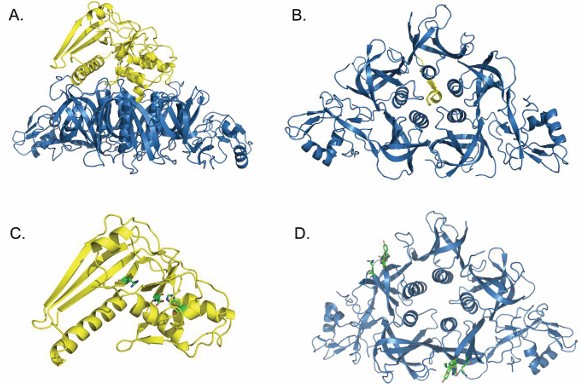 Figure 1. The structure of pertussis toxin. (Source: Locht C, et al. 2021)
AB5 Toxin:
Assembly and Secretion:
The A subunit and B pentamer non-covalently associate to form the holotoxin in the periplasmic space of the bacterium. The holotoxin is then transported outside the bacterial cell by a Type IV secretion system.
Part 01
Cellular Entry and Intracellular Trafficking
The B pentamer attaches to sialoglycoproteins on the cell surface. The toxin enters the cell through endocytosis, and is taken on a unique retrograde transport pathway: from the endosomes, through the Golgi, and finally to the ER.
Part 02
Activation and Cytosolic Translocation in the ER
Inside the ER, the binding of ATP will dissociate PTS1 from its interaction with the B pentamer, causing the release of PTS1. Free PTS1 is thermally labile under physiological conditions, which causes it to unfold. The unfolded PTS1 can now be targeted by the cellular ER-associated degradation (ERAD) pathway. ERAD is responsible for moving misfolded proteins from the ER into the cytosol for degradation. PTS1 exploits this pathway to be delivered into the cytosol.
Part 03
Mechanism of Toxicity
Once the active PTS1 reaches the cytosol, it executes its enzymatic function, primarily through:
ADP-ribosylation;
Loss of function of its target;
Disruption of cellular signaling;
Cell-type-specific effects.
 Figure 2. Schematic depiction of the cellular uptake and mode of action of PT. (Source: Ernst K. 2022)
Creative Diagnostics provides high-quality antibodies, antigens, and various reagents related to Bordetella pertussis to support your research and development needs.
These are the first-line drugs of choice.
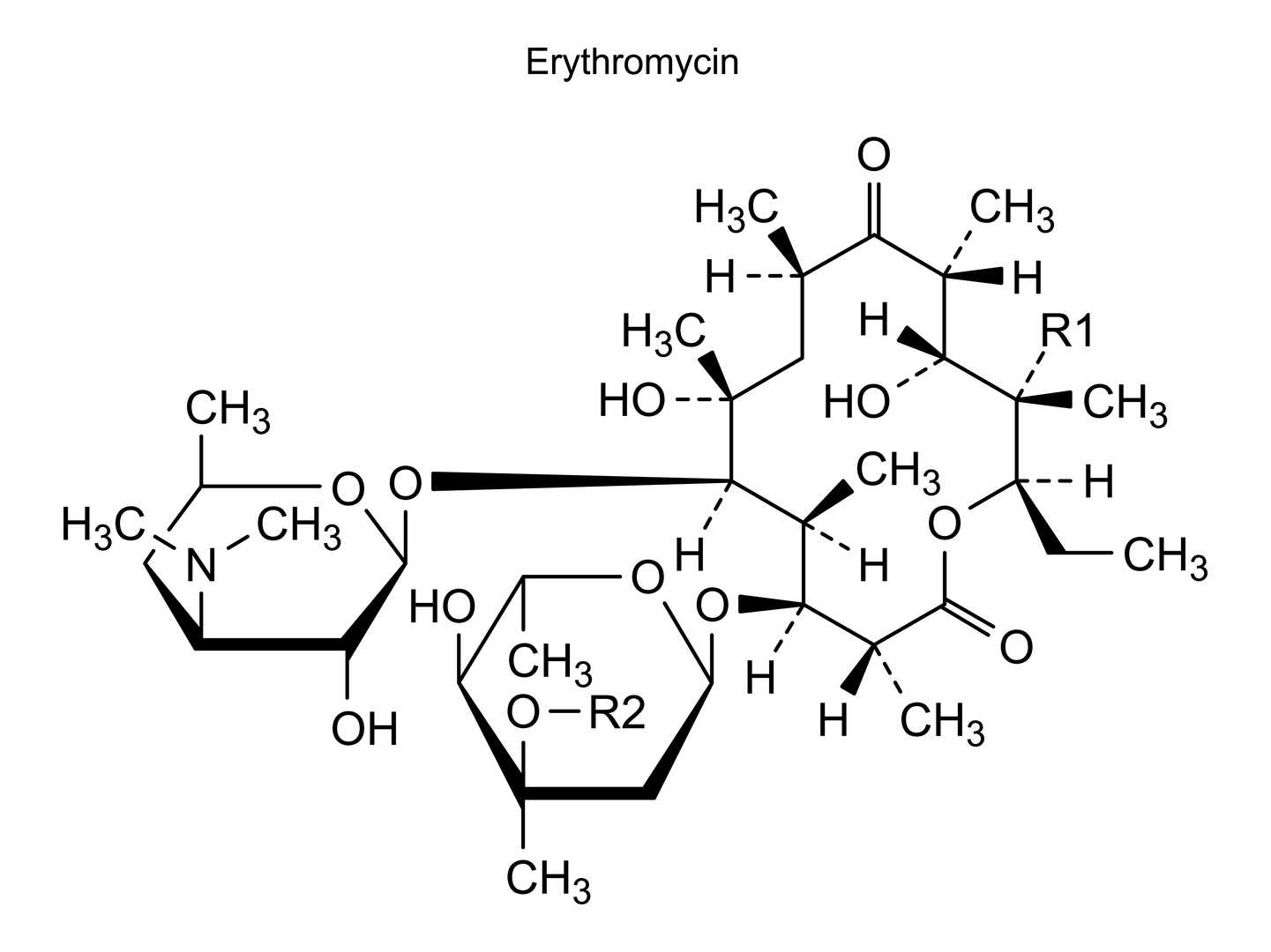
Traditional Option: Erythromycin, but note the following:

Modern Preferred Choices:
For patients who cannot tolerate macrolides, Trimethoprim-Sulfamethoxazole can be used.
References
Loading ......
