
Inquiry Basket
 Loading ......
Type 1 diabetes mellitus (T1DM) is an organ-specific autoimmune disease characterized by pancreatic β-cell destruction, driven by the activation of lymphocytes against pancreatic β-cells. beta cell proteins as autoantigens (AAgs) are recognized by patients' own CD4+ and CD8+ T cells and/or the products of self-reactive B cells, called autoantibodies. Well-established autoantigens (AAgs) include islet cell autoantigen (ICA), insulin, glutamic acid decarboxylase (GAD), tyrosine phosphatase-like proteins insulinoma antigen-2 (IA-2), zinc transporter-8 (ZnT8). Autoantibodies of these AAgs serve as key markers in immune-mediated type 1 diabetes (T1D). Understanding the nature and clinical utility of AAgs is a central focus in diabetes research and has important implications for prediction prior to disease onset, diagnosis, and intervention by restoring immune tolerance.
T1DM results from the destruction of the insulin-producing cells in the pancreas called beta cells by the adaptive immune system. Genetic factors and one or more environmental factors lead to the recognition of beta cell components as autoantigens leading to an autoimmune attack. autoantigens, such as, insulin B chain peptide (11-23) and other components of beta cell secretory granules (GAD 65, IA-2, and transmembrane Zn transporter, et al.) are presented by HLA molecules major histocompatibility complexes (MHC) I and II on antigen presenting cell (APCs) to diabetogenic autoreactive T cells. Autoreactive CD4 T cells stimulate APCs, including B cells that produce high-affinity autoantibodies against beta cells. Autoreactive CD4 T Cells also help diabetogenic CD8+ T cells to acquire cytolytic activity and attack beta cells through the release of cytokines (including TNF-α and IFN-γ, Fas/FASL and perforin/granzyme). Released cytokines also stimulate macrophages and other innate immune cells to further damage beta cells yielding a positive feedback loop with the production of more toxic cytokines to propagate further beta cell destruction.
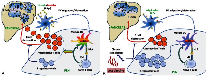 Fig. 1 A. Autoantigens-mediated pancreatic β-cell destruction. B. Antigen-specific immunoregulation. (Han S., et al., 2013)
GAD is a rate-limiting enzyme that catalyzes a decarboxylation reaction to produce the neurotransmitter γ-aminobutyric acid from L-glutamate. There are two isoforms of GAD, GAD65 (585 amino acids), and GAD67 (594 amino acids), both of which are abundant in the central nervous system and pancreatic islets. In pancreatic β-cells, GAD65 and GAD67 are localized in the synaptic-like vesicles and cytosol, respectively. GAD autoantibodies can accurately predict T1D development in combination with other surrogate humoral biomarkers.
Zinc ions are essential for the proper storage, secretion, and action of insulin. The zinc content in the pancreatic beta cell is among the highest in the body. Zinc ions are transported from the cytoplasm to insulin secretory vesicles in pancreatic beta cells by the SLC30A zinc transporter 8 protein (ZnT8). ZnT8 is specifically expressed in pancreatic beta cells and has been identified as a novel T1DM autoimmune target. ZnT8 autoantibodies have been associated with rapid onset of hyperglycaemia and eventual loss of graft function, making them a potentially important biomarker for predicting beta cell loss.
IA-2 is a transmembrane glycoprotein of the protein tyrosine phosphatase (PTP) family that colocalizes with IA-2β/phogrin and is expressed in insulin-secretory granule membranes in pancreatic β-cells. Both polypeptides regulate insulin secretory granule content and pancreatic β-cell growth. IA-2 AAb may allow for better characterization of the risk of progression of T1D, improve staging accuracy of presymptomatic T1DM. IA-2 is likely to be the primary target of humoral autoimmunity against diabetes associated PTP-like autoantigens. IA-2 AAb may allow for better characterization of the risk of progression of T1D, and improve staging accuracy of presymptomatic T1DM.
ICA69 is predominantly expressed in pancreatic islets and neuroendocrine organs. It is a conserved regulator of neuroendocrine secretion. ICA69 is involved in DCV signaling and maturation, and it is recruited to Golgi membranes by activated Rab2.
 Fig. 2 Predominant intracellular distribution of major T1D autoantigens in pancreatic β cells. (Arvan P., et al., 2012)
The clinical applications for AAgs in T1DM AAgs can be used in two settings for immunotherapy of T1DM.
1). Specific detection of autoantibodies against multiple islet AAgs
Specific assays used to detect autoantibodies against multiple islet AAgs are both diagnostic and predictive markers for T1DM. They may help to discriminate T1DM and T2DM, diagnose acute-onset, ketoacidotic diabetes in obese individuals, predict and prevent the development of T1DM, et al.
2). Antigen-specific immunotherapy.
Islet AAgs can be used as therapeutic agents to induce immune tolerance, which has the advantage of focusing treatment on self-reactive T cells without impairing immune responses to unrelated antigens, especially tumor and infectious antigens. Antigen specific immunotherapies have already been applied for preventing and treating T1DM in preclinical models and in patients. Experiments show that the administration of GAD65 can prevent autoimmune destruction of pancreatic beta-cells in experimental animals. GAD-alum was developed and, after necessary safety investigations, was used in a Phase 1 trial in LADA patients with promising results. Although some immune-modulatory agents showed very promising effects in animal models, none have resulted in stable, long-term insulin independence in human diabetic patients. Thus, in order to improve diagnostic and predictive accuracy of islet autoantibodies and preventive and therapeutic efficiency of antigen specific immunotherapy, it is extremely necessary to further study the key AAgs that may initiate and drive disease progression.
References
| Cat. No | Product Name | Expression System | Application | |
| DAGC025 | Recombinant Human ZnT8 | Yeast | ELISA | Inquiry |
| DAG-WT1343 | Recombinant Human ZnT8 | HEK293 cells | ELISA, CLIA, CG | Inquiry |
| DAG-ZNT8I | Recombinant Human ZnT8 | Insect cells | ELISA, WB | Inquiry |
| DAG-ZNT8 | Recombinant Human ZnT8 | E. coli | ELISA, WB | Inquiry |
| DAG-WT1346 | Recombinant Human GAD65 | HEK293 cells | ELISA, CLIA, CG | Inquiry |
| DAGC128U | Recombinant Human GAD65 | Insect cells | ELISA, WB | Inquiry |
| DAGAD6501 | Recombinant Human GAD65 | E. coli | ELISA, WB | Inquiry |
| DAG-WT1345 | Recombinant Human IA2 | HEK293 cells | ELISA, CLIA, CG | Inquiry |
| DAGC269 | Recombinant Human IA2 | Insect cells | ELISA, WB | Inquiry |
| DAG4558 | Recombinant Human IA2 | E. coli | ELISA, WB | Inquiry |
| DAG-WT1344 | Recombinant Human ICA | HEK293 cells | ELISA, CLIA, CG | Inquiry |
| Cat. No | Product Name | Applications | |
| CABT-L4633 | Mouse Anti-Glutamic acid decarboxylase monoclonal antibody, clone 22 | IHC, WB | Inquiry |
| DMABZ008 | Mouse Anti-Human GAD65 monoclonal antibody, clone 255 | WB, IP, FC, RIA | Inquiry |
| CABT-L6004 | Human Anti Human GAD65 monoclonal antibody, clone D22 | ELISA | Inquiry |
| DPABA-4191 | Goat Anti-Human GAD65 polyclonal antibody | WB | Inquiry |
| CABT-L6308 | Human Anti-Human ZnT-8 monoclonal antibody, clone U0 | ELISA | Inquiry |
| CABT-L6307 | Human Anti-Human ZnT-8 monoclonal antibody, clone U3 | ELISA | Inquiry |
| CABT-B8539 | Mouse Anti-Human SLC30A8 (aa 268-359) monoclonal antibody, clone 926140 | ELISA, IHC | Inquiry |
| DPABH-27011 | Rabbit Anti-Human SLC30A8 (internal region) polyclonal antibody | WB | Inquiry |
| DMABZ009 | Mouse Anti-Human IA-2 monoclonal antibody, clone 87G | WB, IP, IHC | Inquiry |
| CABT-L6446Z | Goat Anti-Human IA-2 polyclonal antibody | ELISA, WB | Inquiry |
| CABT-B10442 | Mouse anti-Human ICA1 monoclonal antibody, clone 7H22 | ELISA | Inquiry |
| DPABH-04384 | Rabbit Anti-Human ICA1 (aa 1-223) polyclonal antibody | WB, IHC-P | Inquiry |
Loading ......
