Introduction of Protein-A Colloidal Gold Flow-Through
Immunodiagnostics play a very important role in disease diagnosis. Initial immunodiagnostics were based on the radiolabeling of an antigen or antibody and were limited to a few sophisticated well- equipped labs. With the advent of the enzyme-linked immunosorbent assay (ELISA) and other non-radiolabeled immunoassays, the use of immunodiagnostics became more popular. The major limitations of ELISA are (a) its microplate format which is more suitable for surveillance or for big hospital settings rather than for individual samples, and when inordinate, waiting time may be imposed till enough samples have accumulated, (b) time and trained manpower required for testing, and (c) specialized equipment to determine cutoffs for positive and negative samples. Three different immunoassays are being used for the diagnosis of infectious agents based on membrane matrices, i.e., lateral flow assay (LFA), dot immunobinding assay (DIA) or flow-through assay (FTA), and dipstick assay (DSA). These tests are often referred to as rapid diagnostic tests (RDTs), bedside tests, pen side tests, or point-of-care tests (POCT). Most of the RDTs are semiquantitative or qualitative tests, simply differentiating between positive and negative by the development of a dot or change in color or development of a colored line. These systems are useful in resource- limited laboratories and hospitals and also to take quick decisions on the course of treatment without waiting for the results of time- consuming tests like ELISA or immunoblotting. In all three assays, the principle is the same, i.e., antigen-antibody complex formation and visualization of the complex through color developed from a chromogenic substrate. All these qualitative assays are also less time consuming and can be employed without any need of equipment and training.
Flow-through assay is a rapid test as the total procedure is completed within 3–5 min, is simple to perform, is cost-effective, and can be performed with minimal training without any laboratory equipment. Several studies have reported the use of FTA for the detection of hormones, insecticides, viral antigens, and serum antibodies. Though these tests can be used to detect both antibodies and antigens, sensitivity is more for antibody detection assays, although the detection of antigen is often less sensitive than lateral flow or traditional enzyme immunoassay (EIA) methods. However, FTAs are preferred over lateral flow tests for the simultaneous diagnosis of multiple concurrently occurring pathogens, e.g., multiple rapid HIV/HCV antibody test.
Before developing any FTA system, one needs to decide the analyte to be detected, i.e., antigen or antibodies (see Note 1). Among the reagents to be used, one needs to consider the following points before planning for the development of the assay: (a) type of antigens, i.e., proteins (native or recombinant), peptides, and polysaccharides, as well as the purity of the antigen; (b) type of antibodies (monoclonal or polyclonal) and their specificity, affinity, and avidity; (c) the most suitable format for detection, i.e., direct, indirect, competition, inhibition, etc.; and (d) chromogenic substance. Unlike enzyme-based color development, colloidal dye particles are used in FTA (see Note 2). Initially, colloidal gold conjugated antibody or antigen for the detection of antigen or antibody, respectively, was used on a nitrocellulose membrane in dot immunobinding assay. Subsequently, colloidal gold labeled protein A is being used as a probe for the detection of antibodies in serum samples. For immunoblotting, membrane-based matrices are used along with an absorbent material (see Note 3).
The development of an FTA for detection of antibodies to parasites belonging to Taenia solium is described here employing porcine cysticercosis as a model. Antigens in the form of cyst fluid (CF) and whole cyst (WC) antigens of Taenia solium metacestodes coated onto nitrocellulose membrane and test sera are probed with a protein-A colloidal gold conjugate. The sensitivity and specificity of FTA were in agreement with those of ELISA when conducted with CF and WC antigens. The whole procedure could be completed in 3–5 min at room temperature with visualized results of colored spots.
Materials of Protein-A Colloidal Gold Flow-Through
1. Flow-through modules contain an absorbing pad between upper and lower plastic casings (see Note 3).
2. Nitrocellulose membrane, 0.45μm pore size (see Note 3).
3. Wash solution [phosphate-buffered saline—Tween 20 (PBST), pH 7.4]: 0.20 g potassium chloride, 0.20 g potassium dihydrogen orthophosphate, 8.00 g sodium chloride, and 1.16 g disodium hydrogen phosphate are dissolved in 800 ml of distilled water, 5 ml of Tween 20 is added, and the solution is made up to 1,000 ml.
4. Blocking solution: dissolve 5 g of skimmed milk powder (any high-quality powder available in grocery store) in 100 ml of PBST (see Note 4).
 Figure 1. Flow-though device: (a) Upper casing, (b) nitrocellulose membrane, (c) absorbent pad, (d) lower casing.
5. Antigens: cyst fluid antigen (CFA) and whole cyst antigen (WCA). Prepare CFA and WCA as reported (see Note 5).
6. Conjugates: colloidal gold conjugate (see Note 2).
Methods of Protein-A Colloidal Gold Flow-Through
Flow-through assay (FTA) for antibody detection: The procedure can be divided into two components, i.e., antigen coating and testing sera for antibodies. The procedure described here includes the preparation of antigen-coated devices for future use. However, the procedure can be modified to prepare a few devices and use them immediately. Carry out all procedures at room temperature unless specified otherwise. Test principle: In the assay, antibodies in the serum sample are captured by an antigen spotted onto a nitrocellulose membrane mounted on a flow-through device that serves as the antigen capture matrix. The bound antibodies are visualized by the addition of protein-A colloidal gold conjugate, which imparts a pink color to the membrane.
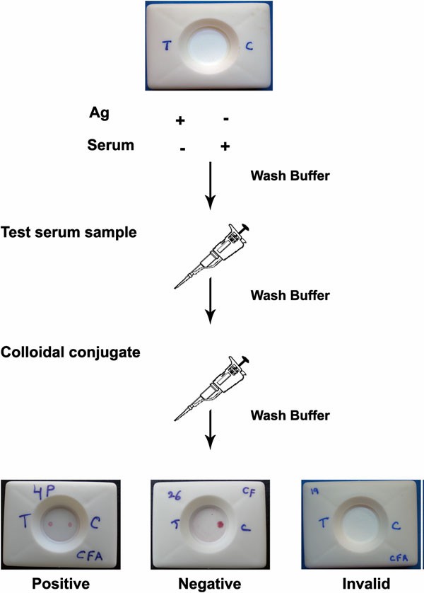 Figure 2. Schematic depiction of flow-through assay.
Antigen Coating
- Place the nitrocellulose membrane on a blotting paper (see Note 6).
- Place 1μl (250 ng/μl) of antigen at one end ("T" side) and 1 μl of pig serum at the opposite end ("C" side) (see Note 7).
- Dry the membrane in an incubator at 37 °C for 1 h or at room temperature overnight (see Note 8).
- Wash the membrane with 200μl of wash buffer and allow wicking through the membrane by capillary action (see Note 9).
- Dip the membranes in blocking buffer, incubate at 4 °C for 1 h, place the membrane on blotting paper, and dry the membrane by incubating at 37 °C for 1 h or at room temperature overnight (see Note 10).
- Place the membrane in flow-through module making sure that the membrane is tightly in contact with the underlying absorbing pad (see Note 6).
Testing Sera for Antibodies
- Add 200 μl of test serum diluted 1:10 in wash buffer and allow it to be absorbed through the membrane (see Note 11).
- Add 200 μl of wash buffer and wait until the buffer is absorbed completely.
- Add 200 μl of protein-A colloidal gold conjugate diluted 1:2 in wash buffer and allow to be absorbed through the membrane (see Note 12).
- Add 200 μl of wash buffer and wait until the buffer is absorbed completely.
- Appearances of two pink dots indicate the presence of antibodies and indicate that the test is positive. Appearance of no color at T dot with pink color at C dot indicates that the result is negative. If no pink color appears at C dot, the test is invalid and the test needs to be repeated (see Note 7).
View ELISA Matched Antibody Pair
Notes of Protein-A Colloidal Gold Flow-Through
- Immunodiagnostics can be basically divided in to two systems, i.e., those that detect the antigen or the etiological agent and those that detect antibodies against the agent. These tests respectively employ specific antibodies or antigens. Antigen detection is more useful for acute diseases (e.g., dengue fever, malaria) and to detect an ongoing infection and can assist clinicians to tailor treatment regimen. This includes the detection of whole organism or parts of it. The identification of antibodies from the patient's serum is more useful for the diagnosis of chronic infections like those caused by hepatitis B and human immunodeficiency viruses. The use of antibody detection assays cannot differentiate the current versus previous infections unless antibody types are detected, e.g., identification of IgM antibodies for the detection of ongoing dengue infection. As a corollary, such tests will be misleading in endemic areas because most of the population shows detectable level of antibodies owing to prior exposure to the specific or a closely related pathogen. For instance, both ELISA and RDT tests for tuberculosis are discouraged for use in endemic areas. On the other hand, antibody detection tests are more useful for epidemiological surveys and are the only assays used for the detection of vaccine induced antibodies.
- In ELISA and EIA, an enzyme is conjugated to the antigen or antibody and the detection is based on the production of color when the substrate is converted to a colored substance by the enzyme. In rapid assays, colloidal gold is conjugated to antibodies or protein A, G, or A/G (though colloidal nonmetals like selenium are available, gold conjugates are more popular; colloidal conjugated protein A and antibodies are commercially available). Proteins A, G, and L are naturally occurring immunoglobulin-binding proteins produced by bacteria. Proteins A and G bind to the Fc portion of antibodies, whereas protein L binds to the light chain of antibodies in the Fab region. All these proteins differ in their antibody-binding specificities and a recombinant protein A/G has broader antibody specificity than either of proteins A or G. The selection of protein A, G, or A/G also depends on the antibody type and species. Since proteins A and G do not or weakly bind IgM, the detection of IgM antibodies is best achieved using anti-IgM antibodies conjugated with colloidal gold. Hence, it is essential to carry out a pilot assay to test the binding of the conjugated protein A/G to the antibodies being used. If a sandwich test is used, where an antigen is sandwiched between two types of antibodies, anti-immunoglobulin conjugates are more preferable than protein A/G conjugates. Different sizes of colloidal gold particles are available for different applications. In general, for immunoblotting assays, colloidal gold particles of >15 nm size are used (20 nm size particles are commercially available).
- Different matrices are available for coating the proteins. Nitrocellulose membranes are considered most suitable for FTAs. Pore size of the membrane plays an important role in coating and assay time. Pore sizes from 0.22 to 0.45μm are generally used. Higher pore sizes are used for other assays like LFA. Protein binding is better with membranes with smaller pore size. In addition, the flow rate is slower allowing enough time for the antibody to bind. Together, higher protein binding and slow flow rate produce higher sensitivity. On the other hand, slow flow rate can also lead to higher background and longer assay times. Different absorbent materials are available for fluid absorption, and the thickness of the absorbent pad and its absorption capacity need to be considered based on the volume of the fluid (sample, wash buffer) or the number of washes being used in the test. In general, pads that can absorb 4 ml of liquid are suitable for FTA. Care should be exercised to keep the absorbent pad in close contact with the membrane for proper flow of the reagents during the assay; hence, the thickness of the pad should be sufficient to keep the pad and membrane in close apposition.
- In FTA, antigens/antibodies are applied to the nitrocellulose membranes as small dots, and binding of the proteins to the membrane is based on non-covalent interactions. Drying of proteins on the membrane results in the binding of the proteins to the membrane. As the assays do not involve prolonged incubation, nonspecific binding of antibodies to the membrane is rarely observed, and hence, most of the manufacturers suggest using the membranes without blocking. In case of high nonspecific background, blocking agents can be used. In general, membranes are dipped in concentrated (1–5 %) protein solutions to block uncoated areas of the membranes. Common proteins used are bovine serum albumin (BSA), skimmed milk powder (nonfat dry milk), and fish gelatin in PBS. It is not advised to use sera for blocking as antibodies present in the sera will produce nonspecific reactivity on the whole membrane.
- The adsorption of proteins to the membrane is based on noncovalent interactions and the antigens or antibodies need to be prepared in coating buffer. The pH of the coating buffer plays an important role in the adsorption to the nitrocellulose membrane. Availability of pKa values of the proteins to be adsorbed will be helpful in deciding the pH of the coating buffer, i.e., the pH of the buffer should be more than the pKa value of the proteins. In general, buffer pH values between 8 and 9 work well with most of the proteins. The source of the proteins also plays an important role.
- The antigen-coated membranes can either be used immediately or stored and used later. Antigen coating is the most time-consuming step in the assembly of the flow-through devices. In general, kits are prepared in bulk and stored for future use. For one-time usage, instead of using blotting paper, the membrane can be placed directly over the absorbing pad in the FTA module.
-
The serum will be coated on the control (C) side of the strip. This will act as control for protein-A colloidal gold conjugate binding to antibodies. Colloidal gold-conjugated protein A will bind to this dot and a pink color is produced. The C dot should turn to pink once colloidal gold-conjugated protein A is added to the test device. No color development at the C dot is an indication of test failure and the test needs to be repeated. Test antigen (T) will be placed on the other side of the strip. Care should be taken to place the C and T dots at least 0.5 cm apart. Specific antibodies binding to the antigen are then bound by a colloidal gold conjugate, developing into a pink dot in case of positive test, and no color development is considered negative for specific antibodies.
View all Positive Control
8. If an incubator is not available for drying the flow-through modules, drying can be achieved by overnight incubation at room temperature. Care should be exercised to dry the membrane completely. The proteins bind to the membrane by noncovalent interactions and stability of the bound proteins will be more when the membrane is completely dried.
9. Washing of the membrane will remove the unbound antigen.
10. This step can be avoided when background is not a problem (see Note 4).
11. The dilution of sera to be used is dictated by the quantity of the serum available to test. An initial dilution of 1:5 is suggested. However, if background is an issue, higher dilutions can then be tested. During our preliminary experiments, 1 in 100 serum dilutions provided clear pink-colored dots, but lightly infected pig (on meat inspection) serum samples gave very faint dots. Hence, further dilutions were made and finally 1 in 10 dilutions was found to be optimum even for light infections and it makes the test reagent conservative.
12. Most of the manufacturers supply colloidal gold conjugates in concentrated form and working concentration of conjugate needs to be determined empirically. Dilutions ranging from two to tenfold should be tested for each batch of the colloidal gold conjugate being used.

 Loading ......
