
Inquiry Basket
 Loading ......
West Nile Virus (WNV) is a single-stranded positive-sense RNA virus transmitted by mosquitoes as vectors and belongs to the Flaviviridae family. When WNV enters the body through the bite of an infected mosquito it replicates in various cells and produces viremia. Most people are infected with no apparent symptoms, but some populations still experience clinical symptoms that can range in severity from mild fever to severe neuroinvasive disease with high morbidity and mortality. Since its introduction to the United States in 1999, WNV has become the common mosquito-borne virus in North America. Since its discovery, the virus has spread to most parts of the globe and is now considered the important agent of viral encephalitis worldwide.
WNV particles are spherical and about 50nm in diameter. They are icosahedral nucleocapsids wrapped in a layer of lipid. The genomic length is about 11kb, which contains an open reading frame (ORF) and an untranslated region (UTRs) at both ends. Like other flaviviruses, there is no polyadenosine tail at the 5' end of the WNV genome. The UTR of the viral RNA genome facilitates replication, transcription, translation, and packaging, and WNV RNA is translated into a polyprotein that is cleaved into three structural proteins (capsid, envelope, and premembrane) and seven nonstructural proteins (NS1, NS2A, NS2B, NS3, NS4A, NS4B, and NS5) in the presence of cellular and viral proteases.
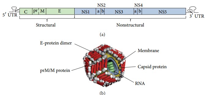 Figure 1. WNV genome organization and virion composition
(Source: Chancey C, et al. 2015)
WNV is transmitted circularly by mosquito-bird-mosquito transmission chain in nature. Different species of Culex mosquitoes are the main vectors of MNV, with Culex tarsalis and C. pipiens considered the main vectors in the western and eastern United States, respectively, and Turdus migratorius being the most important maintenance and transmission host for WNV in the United States. When a mosquito is infected with WNV, the virus infects midgut epithelial cells and begins to replicate before circulating into the mosquito's salivary glands. When an infected mosquito bites a human, the virus inoculates the host's skin, replicates in a variety of these cells, such as neutrophils, macrophages, and keratinocytes, and produces viremia. Since the viral load in human blood is not as high as in birds and is not sufficient to transmit to another mosquito, it is considered the terminal host in the WNV transmission cycle. In addition to mosquito bites, blood transfusions, organ transplants, breastfeeding and laboratory infections may also spread WNV.
WNV infection is a major public health problem in the United States, where the virus has become endemic with recurrent outbreaks for more than a decade.
WNV was first isolated in Uganda in 1937 and has since become endemic in many parts of Africa, Europe, Asia, Australia, and the Middle East. Despite its global distribution, WNV was not detected in the Americas at that time. When health workers observed an unusual spike in unexplained cases of human encephalitis at the same time that bird mortality was very high in New York City in the summer of 1999, it was suspected that it was caused by Saint Louis encephalitis virus (SLEV), a mosquito-borne flavivirus endemic to the Americas that can also cause human encephalitis. However, further testing revealed that the 1999 outbreak of encephalitis was caused by a virus never observed in the Americas - WNV. Phylogenetic comparison of sequences of the virus isolated from New York in 1999 with sequences of WNV from around the world revealed that the virus was most closely related to WNV isolated from the brain of a dead goose found in Israel in 1998, suggesting that the virus may have originated in the Middle East and was most likely endemic in the U.S. for a period prior to the outbreaks first detected in 1999.
 Figure 2. Emergence of WNV in New York.
(Source: Hadfield J, et al. 2019)
This strain of WNV, named WNV NY99, also caused human disease in New Jersey and Connecticut through the summer of 2000, with 21 cases reported, including 19 cases of WNND, resulting in two deaths. In contrast to the pattern of outbreaks in Europe and Africa, where outbreaks were inactive for several years, WNV continued to spread in the United States after its introduction. In the summer of 2001, a total of 66 cases (64 WNND) were reported in 10 states, with 9 deaths. In 2002, 4,156 cases were reported in 40 states, of which 70.9% (2,946 cases) were categorized as WNND, resulting in 284 deaths. In 2003, thanks to the Ministry of Health's outreach to raise awareness among the medical community, the number of WNND cases dropped to 2,866 that year, accounting for 29% of reported cases. At the same time as the 2002-2003 outbreak, a new virus genotype called WN02 replaced the original NY99 virus genotype; the new genotype was observed to be more efficiently transmitted in Culex pipiens and Culex tarsalis mosquitoes than the NY99 genotype. A large outbreak in the U.S. in 2006 resulted in 177 deaths and 1,495 cases of WNND out of a total of 4,260 cases, and another large outbreak of WNV in 2012 resulted in 2,873 cases of WNND in the U.S. and set a record for the highest number of reported deaths in a single WNV season. From 1999 through 2013, 39,557 cases of WNV were reported in the United States, of which 17,381 were WNND cases, resulting in 1,667 deaths, an average of 111.1 deaths per year. Because a proportion of mild cases go unsupervised, epidemiologic estimates suggest that each case of WNND results in 150 to 350 human infections, resulting in 2.6 to 6.1 million people in the United States being infected with WNV over the past 14 years.
 Figure 3. Number of neuroinvasive and non-neuroinvasive West Nile virus cases and infections reported to CDC in the United States, 1999-2013
(Source: Chancey C, et al. 2015)
The rapid spread of WNV in the American continent can be revealed by genomics and phylogenetic analysis. Following the first outbreak in New York in 1999, surveillance of mosquitoes and birds showed that WNV spread along the east coast, reaching Florida in 2001 and westward to the Rocky Mountains and the Pacific Northwest (Washington State) in 2002. Finally, the discovery of WNV in Southern California in 2003 marked the successful spread of WNV in the continental United States. Viral genome data show that WNV is spreading faster in the United States than surveillance data suggest. Combining spatial and phylogenetic data, it is estimated that in the first few years (1999-2003), WNV moved from the east coast to the west coast at an average annual rate of about 1000 km. In addition, when WNV was first detected in New York, the virus had spread to neighboring states and then reached parts of the Midwest and southeast in 2000. The data also show that WNV appeared in Texas as early as 2001 and in California in 2002, a year earlier than the local monitoring system detected. Between 2001 and 2002, the rapid geographical expansion of WNV was consistent with a significant increase in viral genetic diversity and a sharp jump in human cases.
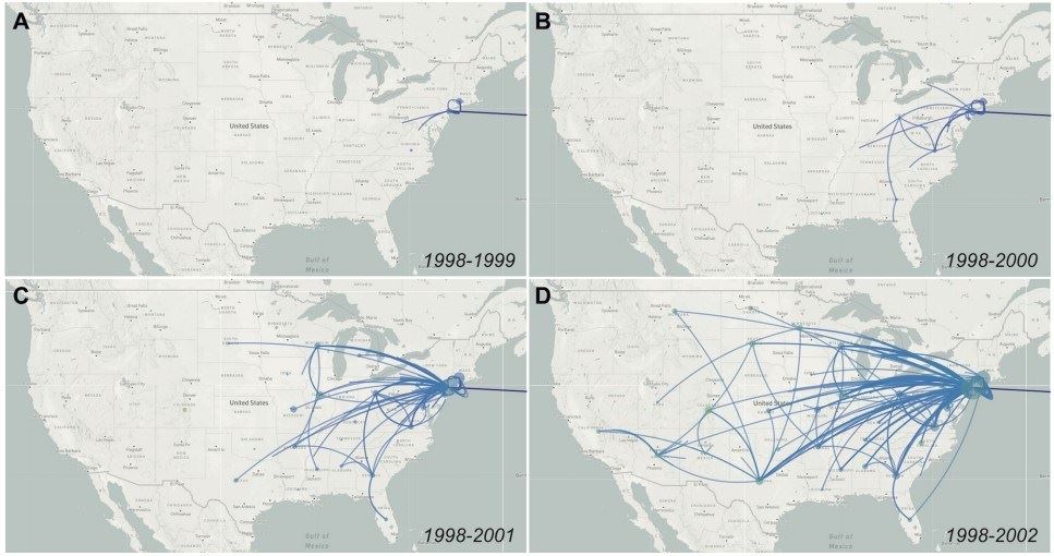 Figure 4. Genomics reveals rapid spread of WNV across the continent.
(Source: Hadfield J, et al. 2019)
WNV infections have become one of the major public health events in the U.S. CDC and state health departments have worked together to establish ArboNET, a National Arbovirus Surveillance System to collect data on arbovirus infections in humans, veterinary animals, mosquitoes, dead birds, and sentinel animals. The current ArboNET collection of MNV infections in the U.S. in 2023 has a total of 2,406 cases reported in 47 states, including 1,599 cases of WNND.
 Figure 5. West Nile virus human disease cases reported by state of residence, 2023
(Source: CDC's ArboNET system)
Clinical Outcomes of Human Infections
Seroepidemiological studies show that 20% of people infected with WNV virus suffer from mild diseases, and one in 150 people suffer from neuroinvasive diseases caused by WNV (WNND). Then, combined with the results of nucleic acid detection, it was found that one out of every 244 to 353 cases of infection would develop into WNND. Between 50%-71% of patients with WNND develop encephalitis, 15%-35% meningitis, and 3%-19% acute flaccid paralysis, with severe case fatality rates for patients with encephalitis ranging from 3% to 19%.
References
| Target | Cat. No. | Product Name | Size | Species | Application | Detection Sample | |
| WNV | DEIAY10297 | West Nile virus NS1 Antigen ELISA Development Kit | 96T | Human | Quantitative | Cell culture supernatants, serum, other biological samples | Inquiry |
| DEIA1982M | West Nile IgM ELISA Kit | 96T | Human | Quantitative and qualitative | Plasma, Serum | Inquiry | |
| DEIA1982 | West Nile IgG ELISA Kit | 96T | Human | Quantitative and qualitative | Plasma, Serum | Inquiry | |
| DEIA-BY005 | Anti-West Nile Virus ELISA Kit (IgG) | 96T | Human | Semiquantitative or quantitative | Plasma, Serum | Inquiry | |
| DEIA-BY005M | Anti-West Nile Virus ELISA Kit (IgM) | 96T | Human | Semiquantitative | Plasma, Serum | Inquiry |
| Target | Cat. No. | Product Name | Expression System | Tag/Conjugate | Application | |
| WNV | DAGA-3052 | Native West Nile Virus antigen(Strain 385-99 (New York)) | N/A | Unconjugated | ELISA | Inquiry |
| DAGA-3053 | Native West Nile Virus antigen(Strain B956 (Uganda)) | N/A | Unconjugated | ELISA | Inquiry | |
| DAGC459 | Inactivated WNV Antigen | N/A | Unconjugated | Inquiry | ||
| WNV Envelope Protein | DAGC742 | Recombinant WNV Envelope Protein (a.a. 295-418) [His] | E.coli | His | ELISA | Inquiry |
| DAG-WT367 | Recombinant West Nile Virus Envelope VLP [His] | HEK293 cells | His | ELISA | Inquiry | |
| DAG566 | WNV Envelope protein [His] | E. coli | His | ELISA, WB, LFIA | Inquiry | |
| WNV NS1 | DAG2333 | Recombinant WNV Nonstructural Protein 1 (a.a. 782-110) [His] | E. coli | His | WB, ELISA | Inquiry |
| DAG2334 | Recombinant WNV Nonstructural Protein 1 [His] | HEK293 cells | His | WB, ELISA | Inquiry | |
| WNV NS3 Protease protein | DAG2605 | Recombinant West Nile Virus NS3 Protease Protein | E. coli | Unconjugated | N/A | Inquiry |
| WNV PreM | DAG-P2517 | Recombinant West Nile Virus preM Protein (a.a. 124 - 259) [His] | E. coli | Unconjugated | WB, SDS-PAGE | Inquiry |
Loading ......
