
Inquiry Basket
 Loading ......
All cells secrete various types of membrane vesicles, with the exception of specialized cells that secrete hormones or neurotransmitters that release secretory vesicles, known as extracellular vesicles (EVs). Initially EVs were thought to be a means for cells to remove unwanted substances, as research progressed, researchers discovered that EVs carry information about a variety of substances, in a rich variety of types, including proteins, lipids, and nucleic acids, which can respond to the state and function of the parental cell. EVs are involved in cell-to-cell communication by exchanging components between cells, and they play an important role in both normal cells and in the development of disease. Extracellular vesicles can be broadly categorized into two main groups based on their biogenesis, namely exosomes and microvesicles. Both are also different in size, with exosomes generally being less than 200 nm in diameter and microvesicles up to 1000 nm in diameter.
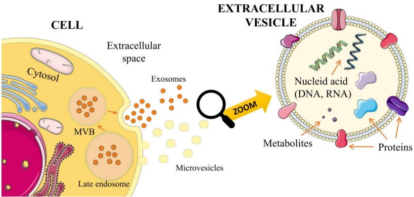 Figure 1. Origin of extracellular vesicles
(Source: Nowak M, et al. 2023)
Exosomes and microvesicles have different modes of biogenesis, with exosomes being produced in the endosomal system as ILVs and secreted outward from the cell via membrane fusion of MVEs, while microvesicles originate from the plasma membrane budding outward. Despite their different biogenesis, there are common intracellular and sorting mechanisms involved.
Exosomes are produced as ILVs in the endosomal lumen during maturation of endosomes into MVEs, a process that involves specific sorting mechanisms. Exosomes are produced as ILVs in the endosomal lumen during maturation of endosomes into MVEs, a process that involves specific sorting mechanisms. The sorting mechanism first sorts the cargo onto microdomains at the limiting membrane of MVEs, which subsequently bud inward and divide into small membrane vesicles containing some cytosol. The endosomal sorting complexes required for transport (ESCRT) is also involved, and exosome biogenesis can be categorized into ESCRT-dependent and ESCRT-independent pathways based on whether they are involved in exosome formation. In the ESCRT-dependent pathway, ESCRT-0 and ESCRT-I subunits aggregate ubiquitinated transmembrane cargoes onto the microdomains of MVE and recruit ESCRT-III subcomplexes via ESCRTII for subsequent budding and fission. Independent of ESCRT pathway, syntenin and ALIX replace the function of ESCRT and participate in cargo sorting and vesicle formation together with other biomolecules including tetraspanins and ceramide.
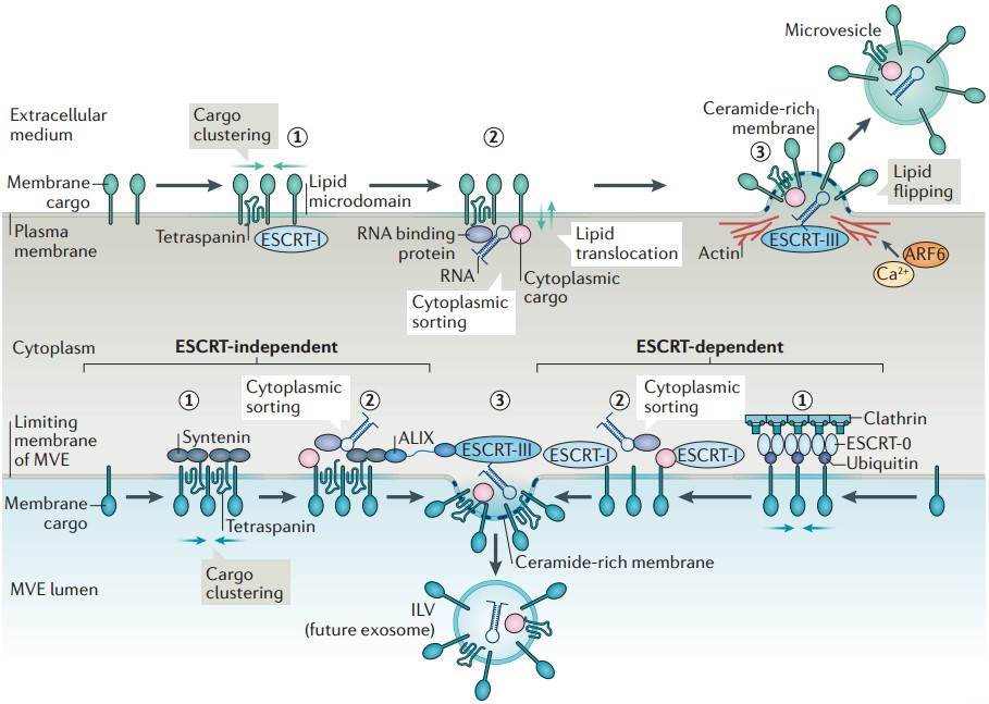 Figure 2. Biogenesis of extracellular vesicles
(Source: van Niel G, et al. 2018)
In the normal circulation, a large number of extracellular vesicles are derived from platelets or megakaryocytes, with a relatively small percentage secreted by the remaining cells. Extracellular vesicles contain proteins, metabolites, and nucleic acids that reflect the origin and function of the cell, at present, the function of extracellular vesicles in blood circulation is still being studied. Extracellular vesicles may play a role in intercellular communication through the transfer of protein and RNA, affecting systemic processes such as immune function and inflammation, as well as a series of disease and organ-specific processes. In view of their importance in intercellular signal transduction and intercellular communication, researchers are more and more interested in their potential role as non-invasive biomarkers for disease detection and prognosis. Extracellular vesicles are considered to be functional biomarkers in some diseases.
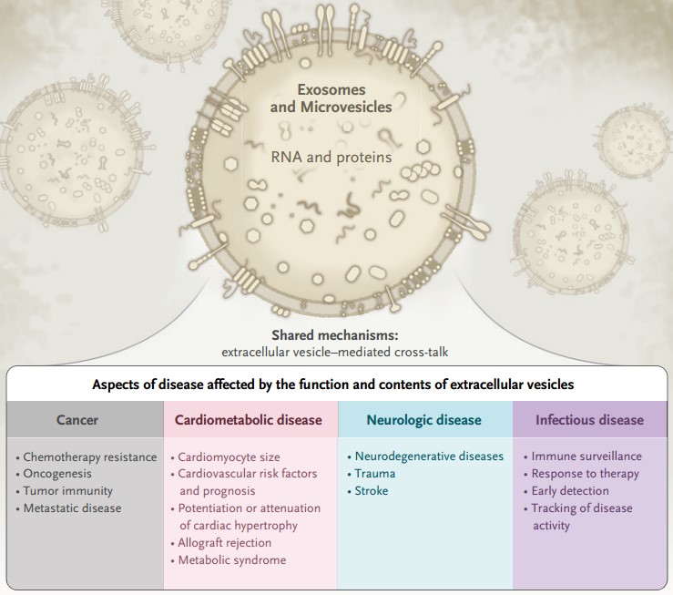 Figure 3. Possible role of extracellular vesicles in the diagnosis and prognosis of various diseases
(Source: Shah R, et al. 2018)
In the treatment of cancer, cancer cells can expel chemotherapeutic drugs from the cells through extracellular vesicles. In ovarian cancer, extracellular vesicles may mediate the interaction between stromal tissue and cancer cells through microRNA 21, thus enhancing the resistance to chemotherapeutic drugs. EVs may also participate in metastasis by carrying molecules involved in epithelial-mesenchymal transition or molecules that prepare the target tissue for metastasis. Based on the fact that extracellular vesicles carry information about their parental cells, the researchers isolated extracellular vesicles from the plasma of patients with acute myeloid leukemia to assess whether they alter the expression of molecules important in immune cell function. These findings relate to the role of EVs in different processes of carcinogenesis and treatment response, and suggest that they may play a role in specific aspects of cancer.
The function of extracellular vesicles in cardiovascular and metabolic diseases has the same characteristics as their role in cancer. For example, angiotensin II induces cardiac fibroblasts to release extracellular vesicles, which can promote cardiac hypertrophy by altering gene expression in cardiomyocytes. In humans, some cardiovascular diseases lead to an increase in circulating extracellular vesicles. Plasma circulating exosome concentrations are proportional to circulating cardiac troponin levels and increase within 24 to 48 hours after coronary artery bypass grafting. The number of circulating microparticles is associated with cardiovascular disease risk factors and long-term cardiac prognosis. Protein studies of circulating extracellular vesicles in heart transplant recipients have shown that the presence of a small number of proteins (some of which involve immune pathways) can identify patients with acute allograft rejection.
Studies have found that extracellular vesicles play a role in neurological diseases. In the model of traumatic brain injury, the increase of miR124 in extracellular vesicles derived from microglia is related to the decrease of inflammation and the improvement of regeneration after injury. In the stroke model, the miR133b in the extracellular vesicles derived from stromal cells may be related to the improvement of neural structure. EVs have also been associated with neurocognitive disorders, and researchers have found that altered expression of key proteins in plasma extracellular vesicles that are directly involved in the physiological functioning of synapses is positively associated with cognitive dysfunction in patients with Alzheimer's disease, and can predict high risk for Alzheimer's disease several years before clinical diagnosis.
Viruses can also use the mechanism of extracellular vesicles for a variety of purposes, including enhancing infectivity and evading the immune system. Extracellular vesicles extracted from hepatoma cells infected with hepatitis C virus in vitro contain genetic information and proteins, which can promote infection without active interaction between virus and target cells, and may escape antibody-mediated immune clearance. In patients infected with human immunodeficiency virus type 1, the size and number of circulating extracellular vesicles are inversely proportional to the ratio of CD4 and CD8 T cells.
Extracellular vesicles can block the infectivity of HIV, regulate immune response, and transmit anti-HIV factors to target cells. Some researchers have found that cells treated with EVs expressing vesicular stomatitis virus glycoprotein can reduce the copy number of HIV proprotein and viral protein Nef. In addition, EVs from other body fluids are also anti-HIV, for example, EVs isolated from healthy breast milk are protective in vitro and EVs isolated from vaginal secretions block the post-entry step of HIV in vitro. These evidences suggest that extracellular vesicles can be used as a new solution for combined antiretroviral therapy and have potential applications in the treatment of HIV infection.
Extracellular vesicles also show therapeutic potential in cancer. EVs packaged with paclitaxel has strong anti-proliferation activity on human pancreatic cancer cells. Macrophage-derived EVs carrying paclitaxel have a high anticancer effect in a mouse lung metastasis model. Red blood cell-derived EVs (RBC-EVs) loaded with doxorubicin or sorafenib enhanced therapeutic efficacy in a mouse model of in situ hepatocellular carcinoma through a macrophage-dependent mechanism and did not show systemic toxicity, in contrast to conventional doses of doxorubicin or sorafenib that showed systemic toxicity at therapeutically effective doses. In clinical trials, injection of EVs derived from dendritic cells into patients with melanoma and non-small cell lung cancer (NSCLC) can appropriately activate T cells, enhance the anti-tumor response of NK cells, and achieve better progression-free survival in patients with advanced unresectable NSCLC. However, due to the complexity of tumor microenvironment, it is difficult for cancer patients to obtain efficient anti-tumor effect. At present, they prefer to improve the efficacy through engineered extracellular vesicles.
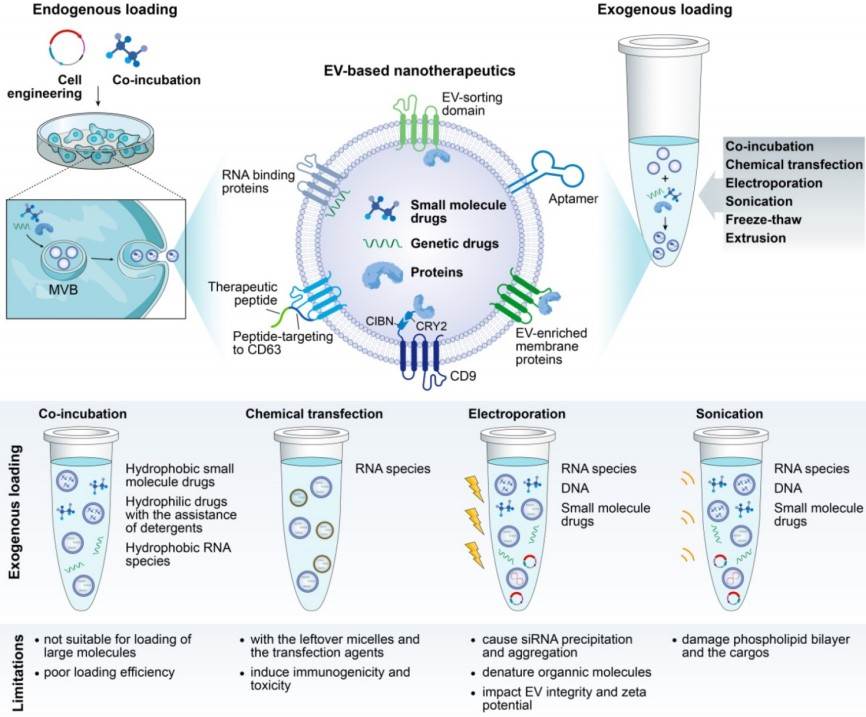 Figure 4. Illustration of cargo-loading techniques to produce EV-based nanotherapeutics
(Source: Tang TT, et al. 2020)
References
| Target | Cat. No. | Product Name | Expression System | Tag/Conjugate | Application | |
| Exosome | DAG-WT1158 | Purified exosomes from MSC | MSC from Adipose tissue | N/A | Calibration, Control, Flow cytometry, EM | Inquiry |
| DAGA-987 | Purified exosomes from plasma of healthy donors | Plasma of healthy donors | Unconjugated | Calibration, Control, Flow cytometry, EM | Inquiry | |
| DAGA-988 | Purified exosomes from urine of healthy donors | Urine of healthy donors | Unconjugated | Calibration, Control, Flow cytometry, EM | Inquiry | |
| DAGA-989 | Purified exosomes from COLO1 cell culture supernatant | Human Colon Carcinoma cell line COLO1 | Unconjugated | Calibration, Control, Flow cytometry, EM | Inquiry | |
| DAGA-993 | Purified exosomes from U87 MG cell culture supernatant | Human Glioblastoma-Astrocytoma cell line U87 MG | Unconjugated | Calibration, Control, Flow cytometry, EM | Inquiry | |
| DAGA-994 | Purified exosomes from SK-N-SH cell culture supernatant | Human Neuroblastoma cell line SK-N-SH | Unconjugated | Calibration, Control, Flow cytometry, EM | Inquiry | |
| DAGA-995 | Purified exosomes from PC3 cell culture supernatant | Human Prostate adenocarcinoma grade IV cell line PC3 | Unconjugated | Calibration, Control, Flow cytometry, EM | Inquiry | |
| DAGA-996 | Purified exosomes from BPH-1 cell culture supernatant | Human being prostatic hyperplasia cell line BPH-1 | Unconjugated | Calibration, Control, Flow cytometry, EM | Inquiry | |
| DAGA-997 | Purified exosomes from DAUDI cell culture supernatant | Human Burkitt Lymphoma cell line DAUDI | Unconjugated | Calibration, Control, Flow cytometry, EM | Inquiry | |
| DAGA-1001 | Purified exosomes from serum of healthy donors | Serum of healthy donors | Unconjugated | Calibration, Control, Flow cytometry, EM | Inquiry | |
| DAGA-1002 | Purified exosomes from saliva of healthy donors | Saliva of healthy donors | Unconjugated | Calibration, Control, Flow cytometry, EM | Inquiry | |
| DAGA-1003 | Fluorescent labeled Exosome Standards | N/A | FITC | Fluorescence microscopy, Flow cytometry, EM | Inquiry |
| Target | Cat. No. | Product Name | Size | Species | Application | Detection Sample | |
| PLVAP | DEIA-FN1144 | Human PLVAP (Plasmalemma Vesicle Associated Protein) ELISA Kit | 96T | Quantitative | Serum, plasma, cell culture supernatants, tissue homogenate | Inquiry | |
| VAPB | DEIA-LL279 | Human Vesicle-associated membrane protein-associated protein B/C ELISA Kit | 96T | Human | Quantitative | Serum, plasma, tissue homogenates and other biological fluids | Inquiry |
Loading ......
