
Inquiry Basket
 Loading ......
The fundamental principle that governs the performance of an immunoassay is the binding of an antibody to a specific antigen to form an exclusive antibody-antigen complex. An antigen or immunogen is any substance that elicits the production of specific antibodies (immune response) in an animal. Any molecule can be considered an immunogen (protein, chemical, nucleic acid, sugar) if it generates an immune response. Molecules that are native to the animal (“self”) are much less likely to induce an immune response than those that are naive or foreign (“non-self”). In general large molecules of high molecular weight function well as immunogens, whereas smaller molecules (drugs, peptides, lipids) require chemical coupling to larger carrier proteins such as keyhole limpet hemocyanin (KLH) or bovine serum albumin (BSA) to generate what is termed a hapten to elicit an immune response. The immune response itself is highly variable and depends on the properties of the antigen, the animal, and method of immunization. Experimentally immunogens are combined with an adjuvant to create an emulsion that is used for immunization to potentiate the immune response. Immunizations are usually repeated over the course of several weeks to maximize the production of specific antibodies circulating in the blood (titer).
The site on the antigen to which a complementary antibody specifically binds is termed an epitope. Antibody-epitope binding may be continuous or linear such as sequence string of amino acids or discontinuous or conformational where binding depends on the three-dimensional shape. Antibody-antigen binding requires that the epitope be available and in the right shape. A multitude of factors can influence epitope availability that include aggregation, masking, chemical fixation or reduction, and changes in pH that may impede or facilitate the non-covalent antibody-antigen binding. Antibody affinity describes the strength of interaction with the antigen at a single antigenic site. All antibody-antigen interactions are reversible governed by thermodynamic principles that are described by an affinity constant (K A). Affinity constants for antibody- antigen binding vary widely and are influenced by temperature, pH, and solvent. Avidity is a measure of the overall stability of the antibody-antigen complex and is mediated by affinity, valence of both antigen and antibody, and the organization of the interacting parts. Together these factors determine the specificity and the probability that a particular antibody will interact with a specific antigen epitope. Cross-reactivity of an antibody refers to binding of an antibody to epitopes on other antigens caused by low avidity or occurrence of identical or similar epitopes on multiple antigens. Cross-reactivity occurs frequently in antigenic groups where chemical structures or amino acid sequences are evolutionarily conserved.
The antibody is the central component of an immunoassay that is used for the detection of a target antigen or analyte. Antibodies exhibit exquisite antigen-binding selectivity at the molecular level which has been exploited to generate probes for the sensitive detection of target analytes used in diagnostic applications. Understanding the characteristics of antibodies and the methods used in their production, purification, and modification is crucial to their use as molecular recognition tools.
Antibodies are glycoproteins produced by plasma cells that function to bind foreign or non-self molecules. In response to a foreign antigen, the host can produce a diverse array of antibodies that are structurally similar yet unique in their properties. Small differences in amino acid sequence result in diverse and distinct properties that mediate antigen-binding versatility, specificity, and biological activity.
This includes a four-chain structure composed of two identical light chains (23 kDa) and two identical heavy chains (50–70 kDa) as their basic unit. Interchain disulfide bonds hold together the heavy and light chains and the two heavy chains. Intra-chain disulfide bonds also contribute to the structure of each heavy and light chain. Differences and similarities in the amino acid sequence of the heavy and light chain Ig divide these polypeptides into variable (V) and constant (C) regions. The light chain consists of one variable (V L) and one constant domain (C L), whereas the heavy chain consists of one variable (VH) and three or four constant (C H 1 , C H 2 , C H 3 , C H 4 ) domains. A hinge region between C H 1 and C H 2 is defi ned by molecular flexibility and is where the arms of the antibody form the Y. Although depicted as a linear Y-structure, the three-dimensional structural map reveals globular domains shaped by intra-chain disulfide bonds as a space-filled/ribbon model derived from the crystal structure of an IgG1. Carbohydrates are attached to the C H 2 domain in most antibodies but are not limited to this domain.
The variable domains show the most amino acid divergence within three regions called the hypervariable regions or complementarity- determining regions (CDRs) localized in both the heavy and light chains. It is the CDR domains of antibodies that are responsible for antigen-binding specificity. The regions between the CDRs in the V L and V H are called the framework region, and the similarities and differences within these regions, resulting from the product of distinct variable region genes, divide both the heavy and light chains into groups and subgroups. Immunoglobulins are divided into five classes (isotype) based on differences in amino acid sequences in the constant region of the heavy chain. IgG and IgA can be further divided into subclasses by additional differences in their constant region of the heavy chains. Yet all immunoglobulins within a class will have very similar sequences within this constant heavy chain region. Differences in the constant regions of the light chains defi ne two light chain types (kappa and lambda), and further differences in the lambda chain result in identified lambda subtypes.
Fragmentation of antibodies by limited proteolysis can be used to create different functional antibody units. Digestion with papain disrupts the IgG before the interchain heavy chain disulfide bonds resulting in the generation of two identical Fab fragments each composed of the light chain and the V H and C H 1 domains of the heavy chain tethered together by the disulfide bridge. In addition an Fc domain is composed of the C H 2 and C H 3 domains of the two heavy chains held together by disulfide bonds. The Fc domain represents the effector functions of the Ig molecule that normally is involved in modulating immune cell function such as the fixation of complement which results in cell lysis or cell receptor binding. The activation of the antibody effector function normally requires prior antigen binding. The enzyme pepsin cleaves the Ig after the intra-chain disulfide bonds between the two heavy chains generating a F(ab’)2 fragment composed of identical Fablike fragments held together by a disulfide bond. The Fc region of the protein is digested into small peptides and a pFc’ domain. The disulfi de bonds make the Ig sensitive to chemical reducing agents such as dithiothreitol (DTT) and 2- mercaptoethanol (2-ME) and effectively disrupt the inter- and intra-chain disulfide bonds and consequently antigen binding.
Antigen binding is primarily mediated by the CDR regions of the variable domains of both the heavy and light chains (VH and V L) used to create the antibody combining site. Valency refers to this antigen-binding determinant composed of CDRs from both the V H and V L, and thus a single Ig is divalent, composed of two identical antigen-binding determinants. The generation of Fab fragments creates two monovalent antibody fragments, whereas the F(ab’)2 retains a divalent binding characteristic.
Immunoglobulin names are based on class/subclass and type. Unless specifically stated, it should be assumed that an “antibody” is a heterogenous composition that includes a combination of class/subclass and type of immunoglobulin. Consider that each immunoglobulin in the mixture will have different antigen- binding properties and the user will have no knowledge of the ratio or composition of the antibodies present. Antisera or polyclonal antibodies will have this composition reflecting a pool of antibodies directed against an antigen. Monoclonal antibodies have a single defined antibody directed at a single antigen-determining region, and the isotype is usually specified.
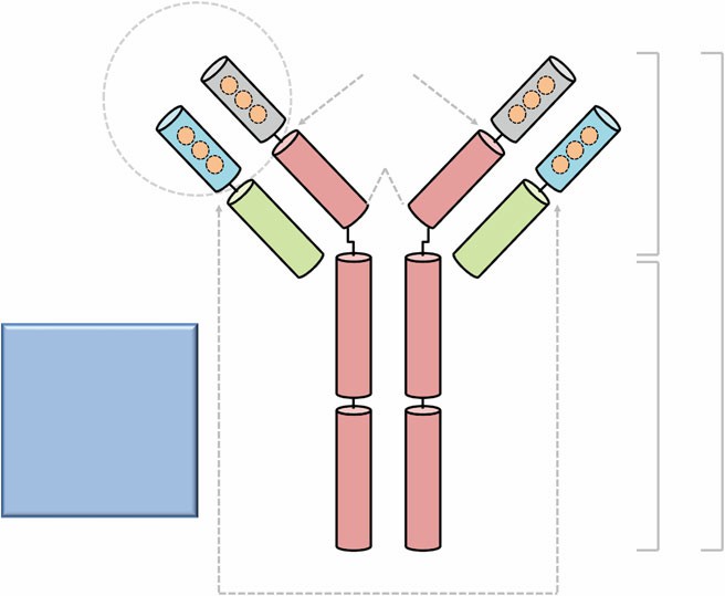 Figure 1. Structural domains of an immunoglobulin.
It is important to differentiate between polyclonal and monoclonal antibodies experimentally in order to maximize their strengths and weaknesses for their application in immunoassays. Many high-quality polyclonal antibodies have been generated for use in immunoassays by repeated immunization of animals with a specific immunogen. Serum is harvested during the peak of antibody production that can yield concentrations of 1–10 mg. The resulting polyclonal antiserum contains many different antibodies capable of binding an immunogen with varied affinity and avidity at a multitude of antigenic epitopes (poly meaning many). Use of polyclonal antiserum in an immunoassay can result in multiple antibodies binding at distinct epitopes on a single antigen which can be used to enhance target detection or isolation. This multiplicity provides added tolerance for an immunoassay where small changes in the antigen presentation may not significantly impact overall binding but consequently serves to limit the discrimination potential of an assay. Understanding the nature of the immunogen used to generate the polyclonal serum can be extremely useful in predicting cross-reactivity. Native or recombinant proteins as immunogens have a higher probability of generating polyclonal antiserum with cross-reactivity to other homologous protein family members. Peptide sequences used as immunogens that have been screened by database search to be unique have lower probability of generating cross-reactive antibodies but can generate antibodies to non-native structure or unavailable epitopes in the parent protein resulting in artifact or an inability to bind native protein. Many of these confounding attributes can be minimized by screening and selection processes of antibodies during or after production (see Note 1). For example, species cross-reactivity of polyclonal antiserum can be minimized by pre-absorption of antiserum with a cross-reactive species to deplete the pool of antibodies that would bind undesirably to that particular species. Monoclonal antibodies (mono meaning one) represent a homogenous pool of identical antibodies which bind to a single antigenic epitope with a defined affinity and avidity. The homogeneity of the monoclonal antibody results in a high degree of specificity but often with a limited tolerance for changes in epitope. This allows for detection of subtle molecular changes between and within antigens but also makes the use of monoclonals more vulnerable to changes in assay conditions. Normal serum contains ~10 16 antibodies per milliliter, and these antibodies can be collected from experimental animals to identify, label, or separate molecules and cells. However the variability of antisera combined with its finite quantity can be disadvantageous when building an immunoassay. The advantage of a polyclonal antibody pool is there are numerous antibodies that can be used to bind a given target at different molecular sites. The disadvantage is that some or many of the antibodies in the pool may exhibit poor binding characteristics, whereas the desired antibody/s in the pool may only be present in limited quantity. Moreover polyclonal antibodies represent a limited commodity that will run out, and effort to generate new antisera will invariably result in a different pool of antibodies even with the same immunogen. Furthermore bioconjugation of polyclonal antibodies is confounded by the varied ratio and quantity of each within a given pool resulting in potentially varied results. Monoclonal antibodies have the advantage of being a continuous renewable resource. Thus the characterized properties of a monoclonal antibody will not change over time, an important consideration in developing an immunoassay and technology transfer. Clear properties of a monoclonal antibody can be defined such as binding affinity (KD), isotype, epitope, and CDR domains. As monoclonal antibodies represent a homogenous pool, they are readily conjugated and can be easily characterized.
Conventional methods for IgG fragmentation to a monovalent Fab fragment are carried out using papain digestion and bivalent F(ab’)2 fragment using pepsin digestion. Papain proteolysis produces Fab fragments from all IgG subclasses and species, whereas pepsin is less universal. Alternate F(ab’)2 fragmentation can be accomplished with those IgGs using the enzyme ficin and is particularly useful for mouse IgG1. To achieve IgG fragmentation into monovalent Fab fragments, preliminary test should be used to determine optimal papain conditions (concentration and time) for IgG fragmentation prior to large-scale antibody digestion. Results of digestion should be compared by Coomassie staining following SDS-polyacrylamide gel electrophoresis to determine optimal conditions. Nonreduced IgG migrates to ~150 kDa, Fab fragments ~50 kDa, and Fc fragments ~27 kDa. Under reducing conditions (10 % 2-ME or DTT), IgG will migrate as two bands of ~50 and 25 kDa, Fab fragments will yield a doublet of ~23–25 kDa, and Fc fragments will migrate as a band ~26 kDa.
37 °C incubator or water bath.
Magnetic stir bar and mixer.
Timer.
1.5 mL microfuge tubes.
Centrifuge.
Rocking platform.
P200 pipette and tips.
Transfer pipette.
Purified IgG at 2 mg/mL in PBS.
Phosphate-buffered saline (PBS); pH 7.2.
0.5 M EDTA (disodium salt) stock in PBS.
Freshly prepared 1 M L-cysteine in PBS.
Papain enzyme (lyophilized powder) in freshly prepared PBS digestion buffer stored on ice (<4 h) containing 0.02 M EDTA (disodium salt) and 0.01 1 M L-cysteine.
Freshly prepare 0.3 M iodoacetamide from solid in PBS.
Immobilized protein -A agarose (6 % cross-linked). Gently mix protein-A agarose bead stock by swirling suspension (do not vortex) and transfer defined volume to a fresh tube using a wide-bore pipette. To remove preservatives, wash beads by dilution in PBS 1:5 swirl and then slow centrifugation for 5 min at 900 × g in fixed angle rotor; repeat. Resuspend beads in PBS equivalent to initial volume taken from stock slurry ~50 %:
2-Mercaptoethanol (2-ME).
Reagents and equipment for gel electrophoresis of proteins.
Colloidal Coomassie brilliant blue (G-250). Suspend 0.5 g Coomassie G-250 dye in a glass bottle by dissolving in 494.2 mL ultrapure water with 50 g ammonium sulfate and 5.8 mL phosphoric acid using a stir bar to create a stock solution. Freshly prepare working Coomassie G-250 solution by transferring 24 mL of well mixed stock suspension to 6 mL of methanol. Mix and add enough to completely cover gel.
Loading ......
