
Inquiry Basket
 Loading ......
β2 Glycoprotein I (β2-Glycoprotein I, β2-GP1) is an important plasma protein that is synthesized mainly by the liver. β2-GP1 circulates in the blood in a bound form with plasma lipoproteins such as very low density lipoproteins and high density lipoproteins, so it is also known as apolipoprotein H (ApoH), which has a very important role in triglyceride metabolism. β2-GP1 has anticoagulant and binding properties to various anionic substances, and is a functional protein that dually regulates the complement and coagulation systems.
β2-GP1 is a single-chain glycoprotein consisting of 326 amino acids with a molecular weight of approximately 50 kDa. The nucleotide sequence and amino acid sequence of β2-GP1 were obtained from the cDNA of a human stem cell-derived hepatocellular carcinoma cell line (HepG2), which consists of five domains, of which domains I to IV are composed of 60 amino acids, and each of which contains highly conserved cystine, proline, and tryptophan. Four conserved cysteines are interconnected to form two internal disulfide bonds. Unlike the first four domains, domain V contains six cysteines.
In the free state, the interaction between domains I and V of β2-GP1 forms a closed "O" shape. When encountering the exposed surface of the negative phospholipid, due to the strong affinity of domain V for the negative phospholipid, the closed loop is opened and forms a "J" structure that anchors to the surface of the negative phospholipid. At this point domain I is fully exposed and the "J" type may be the active form of β2-GP1 in plasma. It is currently believed that when β2-GP1 binds to negative substances, its structure is altered and the antigenic determinants hidden within it are exposed and recognized by anti-β2-GP1 antibodies (aβ2-GP1), which in turn form immune complexes. Studies of mutant proteins and in vitro synthesized peptides of β2-GP1 and the resolution of crystalline constructs revealed the presence of a lipid-binding region on domain V of β2-GP1. The structural properties of β2-GP1 allow it to interact with negatively charged phospholipids on cell membranes in the bloodstream, thus playing an important role in coagulation and immune responses.
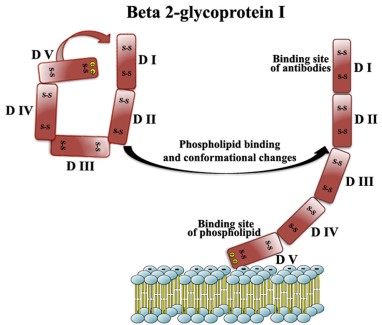 Figure 1. Domains of β2-glycoprotein I
(Source: Sorice M, et al. 2020)
Regulation of anticoagulation
β2-GP1 can exert anticoagulant or procoagulant effects depending on the surrounding environment. As a phospholipid-binding glycoprotein, β2-GP1 acts as a physiological anticoagulant, down-regulating thrombin generation, inhibiting thrombin-induced fibrinogenesis and platelet aggregation, and inhibiting the formation of thrombomodulin complexes, which prolongs the activated partial thromboplastin time (APTT) in normal plasma. However, in some diseases, β2-GP1 exhibits procoagulant effects. β2-GP1 can achieve its procoagulant effects by inhibiting protein C activation and disrupting the crystal shield of anticoagulant annexin V. Clinically, the coexistence of antiphospholipid antibodies (aPL) such as anti-β2-GP1 domain I and anti-phosphatidylserine/prothrombinogen antibodies suggests that the person is at serious risk of thrombosis. Meta-analysis of anti-β2-GP1 antibody classes showed that anti-β2-GP1 IgG was more highly correlated with thrombosis, but in a few diseases anti-β2-GP1 IgM was more correlated with thrombosis.
Platelet regulation by β2-GP1 also affects coagulation to varying degrees. In disease states, the formation of β2-GP1 antigen-antibody complexes can activate platelet receptors, such as apolipoprotein E receptor 2 (ApoER2), G-protein-coupled receptors, and glycoprotein Ⅵ (GP VI), and interact with platelet factor 4 (PF4), thereby affecting platelet activation. Platelet activation expresses tissue factor (TF) and releases other different procoagulant mediators that promote thrombus formation. On the other hand, β2-GP1 inhibits platelet binding to von Willebrand factor (vWF) and interferes with vWF-dependent platelet adhesion, thereby inhibiting thrombosis.
Complement regulation
Some investigators have suggested that β2-GP1 has a role as a cofactor for complement inhibition and can inhibit complement activation through either the classical or bypass pathway. "J"-type β2-GP1 can better bind C3 and provide binding sites for complement factor H, thus mediating the degradation of C3 and cleavage of C3b by complement factorⅠ, which plays an important role in the regulation of complement.
Regulation of angiogenesis
Studies have shown that β2-GP1 modulates angiogenesis in a dose-dependent manner, and that N-β2-GP1 ( nicked β2-GP1) has angiogenic properties at lower concentrations and anti-angiogenic properties at higher concentrations. β2-GP1 down-regulates the expression of vascular endothelial growth factor (VEGF) receptor kinase insert domain receptor/fetal liver kinase 1 (KDR/Flk1) on endothelial cells and prevents the phosphorylation of VEGF downstream effector molecules in the MAPK/ERK and PI3K/Akt/GSK3β pathway.
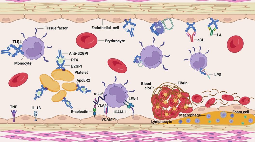 Figure 2. Role of β2-GP1 in injured blood vessels: aPLs consist of aCLs, LA, and anti‐β2-GP1 antibodies
(Source: Li J, et al. 2023)
Modulation of immunity
β2-GP1 has the ability to bind and neutralize lipopolysaccharide (LPS) and has a regulatory role in immunity to infection. In in vitro studies, β2-GP1 was found to inhibit LPS-induced expression of pro-inflammatory factors by monocytes and endothelial cells, negatively modulating inflammation. On the other hand signals released by β2-GP1 and its antibodies can stimulate endothelial cells to release extracellular vesicles, which can stimulate surrounding cells in an autocrine or paracrine manner, and thus have a pro-inflammatory effect. In cellular immunity, β2-GP1 has been found to present β2-GP1-derived peptides to T cells and activate the proliferation and activation of β2-GP1-reactive CD4 T cells, leading to the production of Th1 cytokines, such as IFN-γ predominantly, and the promotion of the production of anti-β2-GP1 antibodies by B cells, resulting in the formation of antigen-antibody complexes, which can lead to the development of diseases such as atherosclerosis.
β2-GP1 is a highly conserved plasma protein that is the major autoantigen in primary systemic lupus erythematosus (SLE) and antiphospholipid syndrome (APS). APS is an autoimmune disease characterized by recurrent thrombosis and pregnancy complications, and testing for β2-GP1 has been included in the laboratory diagnostic criteria for APS since the identification of β2-GP1 as a key autoantigen in APS. Anti-β2-GP1 positivity was significantly higher in SLE patients than in healthy controls.
β2-GP1 binds to a variety of coagulation- and complement-related biomolecules, such as PLs, C3 components, annexin A2, PF4, fibrinogen, and fibronectin. This suggests that β2-GP1 regulates the hemostatic and complement systems, thereby modulating the pathogenesis of APS. On the other hand, apoptotic cells have long been recognized as a source of SLE autoantigens, and the physical interaction of β2-GP1 with these cells provides a "carrier protein"-like connection to a large number of cellular autoantigens. Thus, β2-GP1-reactive T cells have the potential to promote autoantibody production to a wide range of autoantigens expressed by dead cells. It was found that β2-GP1-reactive CD4 T-cell lines from APS and SLE patients primarily recognize epitopes 244-264 within β2-GP1 domain V. They recognize in the context of a variety of HLA class II molecules and exhibit Th0 or Th2-like responses. In contrast, T-cell lines from healthy controls exhibited a Th0 or Th1-like phenotype. Immunization of mice with β2-GP1 and lipopolysaccharide (LPS) resulted in dendritic cells presenting β2-GP1-derived peptides to T cells and activation and proliferation of β2-GP1-reactive CD4 T cells. In the second phase of the response, β2-GP1-reactive CD4 T cells can provide help not only to β2-GP1-specific B cells but also to other autoantigen-specific B cells.
 Figure 3. β2-GP1-reactive T cells promote autoantibody production and pathology in APS and SLE
(Source: Rauch J, et al. 2018)
The "J" type of β2-GP1 exposes the hidden epitope G40-R43 in domain I and allows anti-DI β2-GP1 autoantibodies to bind to it. Antibody recognition against domain I has been found to correlate with APS pathogenesis and clinical manifestations, and therefore detection of anti-DI β2-GP1 antibodies can help in the diagnosis of APS.
Table 1. Overview of assays used for the detection of anti-domain I antibodies (anti-DI)
| Assay | Principle of the method | Exposure of the epitope G40-R43 | Expression of result |
| Two-step ELISA | Antibodies are detected against DI coated on a hydrophilic versus hydrophobic plate by an inhouse ELISA | Yes | Dichotomous values (the ratio of OD on hydrophobic plate/ hydrophilic plate > 2 is positive) |
| Direct ELISA | Antibodies are detected against DI coated on a nickel plate by an in-house ELISA | Not specified | Continuous values (measured in arbitrary units) |
| Competitive inhibition ELISA | The percentage inhibition is calculated when IC50 of DI in solution is used to inhibit antibodies binding to β2GPI coated on a flexible plate by an in-house ELISA | Not specified | The median percentage inhibition of antibodies are calculated |
| Commercial developed ELISA | Antibodies are detected against DI coated on ELISA plates by a commercially developed ELISA | Not specified | Continuous values (measured in arbitrary units) |
(Source: Yin D, et al. 2018)
Reference
| Target | Cat. No. | Product Name | Size | Species Reactivity | Application | Detection Sample | |
| Apo H | DEIA-BJ410 | Human β2-GP1 Ab IgG(β2-Glycoprotein 1 Antibody IgG) ELISA Kit | 96T | Human | Quantitative | Serum, plasma, tissue homogenates and other biological fluids | Inquiry |
| Apo H | DEIA-BJ409 | Human β2-GP1 Ab IgA(β2-Glycoprotein 1 antibody IgA) ELISA Kit | 96T | Human | Quantitative | Serum, plasma, tissue homogenates and other biological fluids | Inquiry |
| Apo H | DEIA-BJ411 | Human β2-GP1 Ab IgM(β2-Glycoprotein 1 Antibody IgM) ELISA Kit | 96T | Human | Quantitative | Serum, plasma, tissue homogenates and other biological fluids | Inquiry |
| APOH | DEIA1862 | ThromboCombo Antibody IgG/IgM ELISA Kit | 96T | Human | Quantitative | Serum, plasma | Inquiry |
| APOH | DEIA1863 | Human Beta-2-Glycoprotein I IgA ELISA Kit | 96T | Human | Quantitative | Serum, plasma | Inquiry |
| APOH | DEIA1814 | Human Anti-Beta2 Glycoprotein I IgG/IgM ELISA Kit | 96T | Human | Qualitative | Serum, plasma | Inquiry |
| APOH | DEIA685 | Human APOH ELISA Kit | 96T | Human | Quantitative | Serum, plasma, cerebrospinal fluid, milk, urine | Inquiry |
| APOH | DEIA05760 | B2GP1 ELISA Kit | 96T | Human | Semi-quantitative, Qualitative | Serum | Inquiry |
| Target | Cat. No. | Product Name | Expression System | Tag/Conjugate | Application | |
| APOH | DAG-WT2778 | Recombinant Human Beta-2-glycoprotein 1 Domain 1 | E. coli | TBD | ELISA | Inquiry |
| APOH | DAGC126 | Native Bovine beta-2-Glycoprotein 1 | Bovien | Unconjugated | SDS-PAGE, WB, ELISA | Inquiry |
| APOH | DAG-T1240 | Beta-2-Glycoprotein 1 Antigen | Inquiry | |||
| APOH | DAG-WT2243 | Recombinant Human ApoH [His] | Mammalian cells | His | Immunoassays | Inquiry |
| APOH | DAG613 | Human Apolipoprotein H [His] | Insect cells | His | WB, ELISA | Inquiry |
| Apo H | DAG4878 | Human β2-Glycoprotein 1 (Apolipoprotein H) [His] | Insect cells | His | WB, ELISA | Inquiry |
| Apo H | DAG4879 | Human β2-Glycoprotein 1 (Apolipoprotein H) | N/A | Unconjugated | ELISA | Inquiry |
| Target | Cat. No. | Product Name | Host | Isotype | Application | |
| APOH | CABT-L511 | Goat anti Human Apolipoprotein-H polyclonal antibody [HRP] | Goat | IgG | IEP, ELISA | Inquiry |
| APOH | DPABH-11734 | Rabbit Anti-Human APOH Polyclonal antibody | Rabbit | IgG | WB, IHC, IF, ELISA | Inquiry |
| APOH | CABT-49300MH | Anti-APOH monoclonal antibody, clone ID2 | Mouse | IgG1 | IHC-Fr, WB | Inquiry |
| APOH | DPAB-DC1686 | Mouse anti-Human APOH polyclonal antibody | Mouse | WB, ELISA | Inquiry | |
| APOH | CABT-L6553Z | Mouse Anti-Human APOH monoclonal antibody, clone II575 [Biotin] | Mouse | IgG1 | WB, ELISA, ELISA(Det) | Inquiry |
Loading ......
