serum, plasma, cell culture supernatant and other biological samples
Species Reactivity
Universal
Detection Method
cELISA
Intended Use
In vitro quantitative determination of Total HCY concentrations in serum, plasma, cell culture supernatant and other biological samples.
Contents of Kit
Note: The liquid reagent bottle contains slightly more reagent than indicated on the label. Please use pipette accurately measure and do proportional dilution.
Storage
2-8°C (for sealed box), please do not freeze! See kit label for expiry date
Performance Characteristics
Perform the stability test for the sealed kit at 37°C and 2-8°C and get relevant data.
Precision
Intra-assay Precision: samples with low, medium and high concentration are tested 20 times on same plate. Inter-assay Precision: samples with low, medium and high concentration are tested 20 times on three different plates.
Detection Range
1.25-80umol/L
Sensitivity
0.75umol/L
General Description
Homocysteine (HCY) is a sulfur-containing amino acid, which is the product of demethylation of methionine. A variety of factors can lead to the accumulation of total homocysteine (total HCY) levels, resulting in hyperhomocysteinemia. The detection of blood homocysteine level is widely used in cardiovascular disease risk assessment and disease prevention and treatment. The normal level of total HCY in human is usually between 5 and 15 μmol/L. Blood homocysteine levels may gradually increase with age. Blood homocysteine levels higher than the normal range may increase the risk of cardiovascular disease, stroke, diabetes and other chronic diseases. More than 80% of HCY in serum or plasma exists in the form of bound or free dimer (HCY-proteins, SAH, HCY-CYS, HCY-HCY, etc.). HCY has no antigenicity. HCY complexes were reduced into monomers under the action of the Antibody Dilution Buffer in this kit, and then S-adenosine homocysteine hydrolase(in Antibody Dilution Buffer) was used to generate Ado HCY for detection of total HCY content indirectly.
Standard Curve
The following assay data are provided for reference, since experimental environment and operation are different. The establishment of standard curve depends on your own assay.
Citations
Publication ()
Have you cited DEIA3576 in a publication? Let us know and earn a reward for your research.
Cdk12 maintains the integrity of adult axons by suppressing actin remodeling
L. N. Townsend, H. Clarke, D. Maddison, K. M. Jones, L. Amadio, A. Jefferson, U. Chughtai, D. M. Bis, S. Züchner, N. D. Allen, W. Van der Goes van Naters, O. M. Peters & G. A. Smith
Cell Death Discovery2023 SepPubMed ID: 37730761Read Article
Applications: ELISA Reactive species: Human
"Abstract: The role of cyclin-dependent kinases (CDKs) that are ubiquitously expressed in the adult nervous system remains unclear. Cdk12 is enriched in terminally differentiated neurons where its conical role in the cell cycle progression is redundant. We find that in adult neurons Cdk12 acts a negative regulator of actin formation, mitochondrial dynamics and neuronal physiology. Cdk12 maintains the size of the axon at sites proximal to the cell body through the transcription of homeostatic enzymes in the 1-carbon by folate pathway which utilize the amino acid homocysteine. Loss of Cdk12 leads to elevated homocysteine and in turn leads to uncontrolled F-actin formation and axonal swelling. Actin remodeling further induces Drp1-dependent fission of mitochondria and the breakdown of axon-soma filtration barrier allowing soma restricted cargos to enter the axon. We demonstrate that Cdk12 is also an essential gene for long-term neuronal survival and loss of this gene causes age-dependent neurodegeneration. Hyperhomocysteinemia, actin changes, and mitochondrial fragmentation are associated with several neurodegenerative conditions such as Alzheimer’s disease and we provide a candidate molecular pathway to link together such pathological events." Article snippet: Fly heads were then homogenized, the supernatant extracted used immediately for a colorimetric human homocysteine ELISA Kit (Creative Diagnostics)
Figure 1. E Actin is more discontinuous in the projections with Hcy treatment. F Actin was enriched in the cell bodies with Hcy treatment.
Effect of arginine:lysine and glycine:methionine intake ratios on dyslipidemia and selected biomarkers implicated in cardiovascular disease: A study with hypercholesterolemic rats
Applications: ELISA Reactive species: Rat
"Abstract: The effect of intake ratios of arginine (Arg): lysine (Lys) and glycine (Gly): methionine (Met) on lipid profile and selected cardiovascular disease markers, was studied, in rats maintained on a hypercholesterolemic diet. The rise in blood cholesterol was countered by 32%, 24%, and 49%, respectively, through increased oral supplementation of Arg, Gly, and Arg+Gly; a corresponding increase in plasma phospholipids at the end of the 8-week study was observed. The elevated plasma cholesterol to phospholipids ratio was countered by 27, 40, and 57%, respectively, through oral supplementation of Arg, Gly, and Arg+Gly. The elevation in hepatic cholesterol was lowered by 18, 29, and 51%, respectively, while phospholipids concentration was concomitantly increased by these amino acids. The elevated cholesterol to phospholipids ratio was, thus, significantly countered in the hypercholesterolemic situation by orally supplemented Arg, Gly, and Arg+Gly. Increased plasma asymmetric dimethylarginine (ADMA) levels, under hypercholesterolemic conditions, were lowered by 12, 15 and 34%, respectively, while plasma symmetric dimethylarginine (SDMA) levels were lowered by 14, 10 and 17%, respectively, with orally supplemented Arg, Gly and Arg+Gly. Only Gly and Arg+Gly decreased plasma homocysteine levels. Total nitric oxide (NO) concentration was considerably increased by Gly supplementation in hypercholesterolemic rats. Thus, altered ratios of Arg:Lys or Gly:Met offered beneficial influence on the lipid profile and plasma levels of ADMA, SDMA and homocysteine in hypercholesterolemic rats. Optimal beneficial effects, among ratios tested, was observed when Arg:Lys and Gly:Met ratios were maintained in ratios of 1:1 and 2:1, respectively." Article snippet: Homocysteine ELISA kitv was procured from Creative Diagnostics (Shirley, NY, USA).
Figure 1. Influence of orally supplemented arginine and glycine on the concentrations of homocysteine and NO in plasma of hypercholesterolemic rats.
Background
Homocysteine (Hcy) is a metabolic intermediate produced by the in vivo demethylation of methionine (Met), which is not present in dietary proteins and does not have to be synthesized endogenously with them. As a sulfhydryl-containing amino acid, Hcy is physiologically essential for processes such as cell cycle progression and maintenance of cellular homeostasis. Hcy metabolism contributes to folate-dependent/folate-independent remethylation to form Met as well as to cysteine formation via the transsulfuration pathway, both of which require vitamin-derived cofactors; vitamin B6 is involved in cysteine synthesis, and vitamins B9, B12, and B2 are involved in the Met synthesis cycle. S-adenosylmethionine (SAM) is a metastable inhibitor of the methylenetetrahydrofolate reductase (MTHFR) reaction and an activator of cystathionine β-synthase (CBS). In the presence of sufficient Met, Hcy produces cysteine via cystathionine β-synthase. In the absence of Met, Hcy undergoes remethylation via N5, N10-methylenetetrahydrofolate reductase to rescue Met. Although Hcy is not directly involved in protein synthesis, its specific functions in folate metabolism and choline catabolism are critical in regulating the effectiveness and function of Met.
Figure 1. Hcy metabolism (Source: Koklesova L, et al. 2021)
Fluctuations in Hcy levels are associated with a wide range of diseases, making Hcy a potent marker of impaired amino acid and protein homeostasis. Four forms of Hcy are present in plasma: the free thiol form (1%), bound to plasma proteins maintaining disulfide bonds (70-80%), bound to other Hcy to form dimeric Hcy, and bound to other thiols (20-30%). The optimal total Hcy concentration (tHcy) in plasma of healthy individuals ranges from 5.0-15.0 μmol/L (high-performance liquid chromatography) or 5.0-12.0 μmol/L (immunoassay). When plasma levels of Hcy exceed the normal concentration range, the patient suffers from hyperhomocysteinemia (HHcy), and a number of risk factors, such as aging, smoking, and oxidative stress, contribute to the severity of HHcy, which can lead to serious conditions, including neurodegenerative diseases, thrombosis, cerebrovascular disease, and cardiovascular disease. Hypohomocysteinemia occurs when plasma Hcy levels fall below the normal concentration range, and low Hcy levels are associated with peripheral neuropathy.
Alternative Names
Homocysteine ELISA Kit tHcy ELISA Kit
References
1. Koklesova L, et al. Homocysteine metabolism as the target for predictive medical approach, disease prevention, prognosis, and treatments tailored to the person. EPMA J. 2021 Nov 11;12(4):477-505
2. Azzini E, et al. Homocysteine: Its Possible Emerging Role in At-Risk Population Groups. Int J Mol Sci. 2020 Feb 20;21(4):1421.
Q: Based on the preliminary HPLC data, the predicted values for hyperhomocysteinemic plasma (EDTA) are ~25 μM in both species, while normal plasma total homocysteine values for rat and mouse are ~5-9 μM. According to the manual on the website, for a 1/dilution ratio, the "amount" in human plasma is ~6-7 μM, while rat is ~14 μM and mouse is ~5 μM. The values in rat plasma are extremely high - does this mean that the "amount" reflects the total concentration in rat and a 1/2 dilution of the other species, or does the kit give better results for some species than others?
A: We speculate that the reasons for this difference may be the following two points: 1. There are differences in the measured values of experimental methods on different platforms. Due to the different detection principles, the quantitative results will be different. This may lead to deviations in the results reported by the same sample under different methods. 2. There are differences in samples. The above data are experimental data obtained by testing limited samples. It represents the typical observations of the kit under specific conditions, but it cannot be completely equivalent to or predict the absolute physiological concentration range of all animal individuals or groups. Individual differences, strains, breeding conditions and other factors may affect the actual measured values. Combining the above factors, we believe that the effect of the hyperhomocysteinemia model can be more effectively evaluated by controlling variables and focusing on relative changes. The comparability of the absolute concentration values of a single sample between different methods or platforms is relatively limited, while the difference trend between treatment groups usually has higher reliability and biological significance.
Customer Reviews
My Review for Total HCY(Homocysteine) ELISA Kit
Creative Diagnostics products are for RESEARCH USE ONLY, please make sure your review is research based.
Required fields are marked with *
Terms and conditions:
We will select high-quality review customers and offer a $30 coupon for your next purchase.
All product reviews must be submitted in the English language.
Creative Diagnostics will not share any personal information of applicants, and all information will be treated with strict confidentiality and will not be sold or disclosed to a third party.
References
Association of methylenetetrahydrofolate reductase C677T gene polymorphism and polycystic ovary syndrome in the South Indian cohort
The polycystic ovarian syndrome is the utmost common endocrinopathy state in women. It is related to both reproductive and metabolic disorders. The MTHFR gene associated with the ovarian follicular action encodes the 5-MTHFR (methylenetetrahydrofolate reductase) enzyme, tangled in folate metabolism. MTHFR gene C677T polymorphism declines the enzyme activity and thus folate deficit and increases the level of homocysteine, which affects the progress of oocytes. Here, we evaluated the association of MTHFR gene C677T polymorphism with Polycystic ovary syndrome (PCOS) in the South Indian cohort of women. About 129 PCOS women with following Rotterdam criteria and 90 women controls were studied. PCR-RFLP technique was carried out on all PCOS women in this study. Dissimilarities in hormone levels in PCOS patients were detected. MTHFR gene polymorphism CC, CT, TT genotype was found to be 74, 16, 9.30% in patients correspondingly. However, in controls, it was 44.4, 24, 31%, respectively. A substantial difference was detected in the genotype frequency distributions among the patients and controls. Also, allele frequency was shown as 82.95% C allele and 17.83% T allele and 56.67%, 43.33% for C, and T allele in controls correspondingly. Our results indicate a possible association and suggest that MTHFR C677T polymorphism can be used as a potential biomarker for PCOS progress in the South Indian women.
Vitamin B-12, Folate and Cognitive Function in Older Adults from Southern Brazil
Objective- To investigate if any combination of serum vitamin B(12)and folate is superior to the use of separate vitamins for identify risk of probable cognitive deficit in older adults from southern Brazil.Methods-We analyzed data from the second wave (2013/14) of a population- and household-based survey. A total of 590 older adults (>= 60 years) were included. Cognitive function was identified by the Mini-Mental State Examination (MMSE). The independent variables were serum levels of vitamin B(12)and folate. The data were adjusted for age, education level, income, smoking status, alcohol intake, food intake, leisure-time physical activity, depression symptoms, chronic diseases, and body mass index.Results- High serum folate levels and their continuous increase were positively associated with cognition. The serum combination of vitamins most found in the study was the excess folate and normal for vitamin B-12, but this combination was not associated with the cognitive function.Conclusion- The combination of folate and B(12)is not high to any of them alone in associating with the probable or absence of cognitive deficit in older adults.

 COA
COA


 Loading ......


 Publication ()
 Figure 1. E Actin is more discontinuous in the projections with Hcy treatment. F Actin was enriched in the cell bodies with Hcy treatment.
 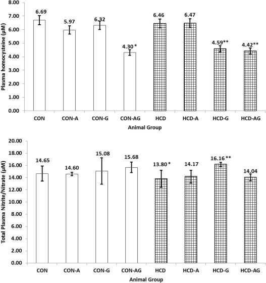 Figure 1. Influence of orally supplemented arginine and glycine on the concentrations of homocysteine and NO in plasma of hypercholesterolemic rats.
  Figure 1. Hcy metabolism
