
Inquiry Basket
 Loading ......
Eukaryotic cells compartmentalize critical cellular functions within individual specialized membrane bound organelles, each tasked with specific functions that are necessary for cell survival—nutrient uptake and processing, gene synthesis, energy generation, protein synthesis, protein transport, waste removal, and so forth. Researchers have utilized the compartmentalization of these functions to ascertain critical clues that may lead to functional determination of a target of interest. As such, much of cell biology has depended on the specific labeling of certain cellular organelles to provide critical information on cellular viability, cellular function, cellular structure, organelle function, organelle localization and number, as well as incorporation within assays that explore the colocalization with a target of interest. A wide range of commercially available dyes and reagents have been created to visualize core cellular structures and organelles, including the plasma membrane, nucleus, endoplasmic reticulum, Golgi apparatus, mitochondria, lysosomes, peroxisomes and cytoskeletal network. However, the extensive breadth of these reagents, which also are available in several spectral variants, has somewhat complicated decisions of which reagent to choose for a particular assay or for the equipment/facilities available to a researcher. Furthermore, each of these reagents has specific characteristics that must be considered within the context of the experimental design and the particular scientific question being addressed. This has created complex algorithms for their use and optimization, especially in multiplexed applications.
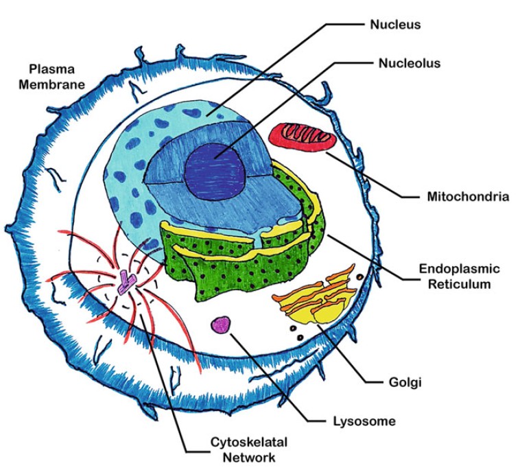 Figure 1. Diagrammatic representation of a eukaryotic cell highlighting some of the specific cellular organelles that are commonly counterstained with organelle specific dyes.
In this introduction, key considerations that should be wellthought- out prior to selecting an organelle dye are outlined. This is followed by a description of each of the core cellular organelles and a summary table of some of the more common commercially available reagents that have historically been employed to label each specific organelle. In addition, within the summary tables are that reagent's associated properties that dictate how and when they can be utilized. Finally, in the ensuing sections complete representative protocols for visualization of each specific core cellular structure or organelle is outlined for a typical reliable example reagent, as well as representative fluorescent micrographs of that labeled organelle. Lastly, notes on potential optimization, troubleshooting or pitfalls are highlighted.
Prior to any decisions on which organelle fluorophore or specific protocol to follow, researchers must first comprehensively consider several key factors related to the researcher's available imaging equipment, the experimental question and design, as well as specifics about the available fluorophores. These questions include the following.
What Specific Fluorescent Imaging Equipment Is Available?
Although large intracellular organelles, such as nuclei, endoplasmic reticulum or cytoskeleton, can be imaged by conventional fluorescent microscopy, smaller sized organelles that are intermingled amongst other organelles and positioned at different levels within the three-dimensional space of a cell may be best cleanly visualized by either deconvolution or confocal microscopy. However, irrespective of their size, given the three-dimensional depth and positioning within a cell, all organelles benefit from compiling a series of optical sections that these types of microscopy provide, while excluding confounding artifacts from out of focus light that may distort the quality of the image. In general, the choice or availability of different instruments also dictate the means of exciting chosen fluorophores or filtering and capturing its emission, the next key consideration in choosing the appropriate fluorophore.
What Excitation and Emission Sources or Filters Are Available on the Microscope?
There is nothing worse than spending hours properly labeling a sample, only to be unable to image it because of not having the matching appropriate lasers to excite or filter sets to capture your chosen probe. For example, while most conventional microscopes have filter sets that can readily image blue fluorophores, there are countless experiences with samples brought to confocal core facilities with nuclei counterstained with DAPI or Hoechst only to discover that there was not a laser line available for exciting and imaging these fluorophores. Therefore, experiments should be designed with organelle probes appropriate to the excitation and emission filter sets available and the specific imaging capabilities of the instrumentation. Careful thought should be taken into what excitation lines and emission filters are available. In cases of instruments with continuous monochromator filter capabilities, one should start by setting the wavelength ranges for the emission profiles of the chosen organelle probe to be captured and narrowing that capture window during optimization. To aid in matching a particular organelle probe to the filters of an available instrument, the peak excitation and emission of representative conjugated organelle probes are included within the organelle summary tables. Although the tables are a small representation of fluorophore conjugates available, where appropriate, the availability of additional fluorophore conjugates with other excitation/emission maximas are noted.
What Are the Spectral Properties of the Fluorophore?
The choice of what fluorophore conjugate to select can sometimes be daunting. There are several significant spectral properties that should be considered in fluorophore selection, which include its excitation, emission, intensity, and photostability. Of these, the primary considerations will usually be based on the fluorophore's excitation and emission profiles (Ex/Em). These profiles should first and foremost be matched to the instrumentation available. Subsequently, the compatibility of the fluorophore Ex/Em within the experimental design should be considered by determining for example, if the selected probes will be multiplexed with other fluorophores. When it comes to a fluorophore's relative intensity upon excitation, fluorophores with higher relative intensities are typically preferred for effective capture and efficient imaging. The choice of a probe's photostability can be dependent upon the experimental design. For many experiments, probes with greater photostability are preferred. However, in fixed samples a probe's photostability can be enhanced during mounting with antifade media. By contrast, in photobleaching experiments, a probe with lower photostability may be desired in order to facilitate bleaching. A final, but not inconsequential, consideration in selection of spectral properties of a particular fluorophore conjugate for an experiment is the autofluorescence potential of either the experimental sample or target organelle. Some tissues or organelles, such as mitochondria and lysosomes, have high levels of autofluorescence that can skew their detection or complicate experimental interpretation. Although autofluorescence can occur across the spectrum, it is most commonly an issue in the 270–400 nm excitation range and the 350–550 nm emission range. Therefore, if autofluorescence is a potential issue with the tissue sample or target organelle, selection of probes with fluorophore conjugates in the far red to near-IR range can alleviate potential issues. The excitation/ emission properties for representative conjugated organelle probes is included within the specific organelle summary tables.
Will the Sample Be Multiplexed for Probing Other Organelles or Proteins?
Although labeling of only an individual organelle within a sample can sometimes address the experimental question, more commonly organelle probes are multiplexed within the context of additional fluorescent probes. Multiplexing of various organelle probes and other targets of interest permits simultaneous detection of multiple markers within a single cell, thereby enabling cellular localization through organelle counterstains, colocalization within specific organelles, and context dependent outcomes within a particular cell or condition. However, the compatibility of each dye's spectral properties must be carefully considered for accurate determination of effects and to prevent misinterpretation of outcomes. Although many fluorescent microscopes today can unmix overlapping spectral properties of dyes, selecting dyes with minimal overlap for the available excitation source and emission filters utilized is the simplest method of ensuring success. Although in the past there was a limited selection of commercially available fluorescent conjugated organelle probes, today's resurgence in interest in organelle probes has fostered the development of numerous probes that match the capability and limitations of current imaging instrumentation. In addition, they provide many spectral variants to enable multiplexing of samples. The better the separation of Ex/Em curve profiles between each of the fluorophores, the higher the probability of a successful well separated multiplexed outcome.
How Does the Fluorophore Function to Label Organelle?
Prior to designing organelle labeling experiments to address a particular experimental question, researchers should have an in depth understanding of the mechanism by which a particular organelle probe labels an organelle. This mechanism can dictate experimental design and whether labeling occurs in live or fixed cells, under certain cellular conditions, only in certain compartments of the organelle or only under defined circumstances. This understanding of how an organelle probe functions can be critical in interpreting results, as well as for troubleshooting or optimizing protocols. For example, due to its mechanism of action in inhibiting protein transport between the ER and Golgi, at higher concentrations use of a brefeldin A conjugated probe for labeling the Golgi apparatus can cause Golgi to ER collapse. Therefore, the staining pattern would appear ER-like rather than Golgi-like. Furthermore, Brefeldin A may affect transport and processing of proteins of interest, thereby changing their localization within the cell. Understanding how each specific probe works can alleviate frustration and misinterpretation of outcomes. Caveats to mechanisms of representative conjugated organelle probes is included within the organelle summary tables.
What Is the Membrane Permeability of the Fluorophore?
The choice of which organelle probe to utilize often is dictated by or dependent upon the experimental design or question being asked. A critical factor in those choices is the membrane permeability of a fluorophore conjugated organelle probe. The plasma membrane of a live cell effectively excludes many dyes from entering and labeling cells, except when cell viability is compromised. Therefore, if the experimental design requires that labeling of an organelle is performed under live cell conditions, then the experimental design dictates that a membrane permeable version of the organelle probe must be utilized. Similarly, if labeling of an organelle is dependent mechanistically upon the cell being live at the time of labeling, then the probe must also be membrane permeable. Conversely, cell impermeant dyes, especially for staining the nucleus, are effectively utilized under live cell conditions to stain only cells that have diminished cell viability as indicated by loss of plasma membrane integrity. Of note, depending on their mechanism of labeling, many membrane permeable organelle dyes can be utilized in both live, as well as fixed and permeabilized cell conditions. The membrane permeability in live cells and their ability to be utilized in fixed cells for representative conjugated organelle probes is included within the organelle summary tables.
Is the Fluorophore Fixable?
Fluorescence-based organelle staining reagents vary in their abilities to maintain signal and be detectable following exposure to cell fixatives. Therefore, the user's experimental design can dictate the choice of which conjugated fluorophores can be utilized dependent on whether that fluorophore is fixable. For example, in live cell labeling experiments where cells will be fixed prior to visualization, organelles must be stained with a fixable fluorophore. However, experiments that more commonly fix, permeabilize and label cells prior to organelle labeling would not be affected by a conjugates ability to maintain fluorescence following fixation. The ability of representative conjugated organelle probes to be fixed is included within the organelle summary tables.
Recently, there has been a renaissance in development of a variety of fluorophores that target specific cell organelles, such as the nucleus, endoplasmic reticulum, Golgi apparatus, mitochondria and lysosomes/ peroxisomes. This resurgence has followed the recognition that the compartmentalization of unique biological processes within each of these organelles can be utilized to understand the biological effects of proteins or other perturbing factors on cell function. In their simplest forms, organelle specific probes are utilized to localize and visualize the organelle within a cell, thereby enabling colocalization studies and some insights into organelle function or cell health. However, more recent advances in organelle probes have resulted in probes that can dynamically assess organelle function in live cells, such as mitochondrial function dyes. Although this Chapter focuses solely on organelle probes for visualization and localization of organelles, many of the described live cell labeling protocols serve as a foundation for the use of cell organelle function dyes. In general, organelle probes are designed to recognize a unique property of the targeted organelle. A fluorochrome is then designed around that process or alternatively, conjugated to that probe to afford target organelle specificity. Because cellular organelles are membrane bound and contained within the cell by the plasma membrane, many organelle probes are also designed to permeate and/or sequester within cellular membranes. This is especially critical for probes designed to work in live cell applications. In these living cells, organelle probes have been utilized to investigate intracellular protein transport, cellular respiration, protein degradation, protein processing, organelle biogenesis, apoptosis, mitosis, and pathogen infection/assembly. By contrast, some organelle probes are cell impermeant, but have been critical in assessing membrane integrity, cell viability, nuclear functions, cytoskeletal structure, and protein transport or retention within and between organelles. In the subsequent sections, a brief description of each target cell organelle with representative organelle probes and their characteristic associated properties are described.
Nucleus
The cell nucleus, derived from Latin meaning kernel or seed, is a double membraned organelle that compartmentalizes and provides protection to the cellular DNA genome. This compartmentalization enriches for machinery required for transcription or DNA replication. It also facilitates both the exclusion and sequestration of transcription factors, enabling highly ordered and regulated expression of genes. Despite having no internal membranous structures, a supporting filament network affords structure and support, while also providing regions within its framework for nonhomogeneous distribution of genomic content. During normal cellular processes, the nucleus may be disassembled or destroyed, either during cell division or due to apoptotic processes. Using specific nuclear stains, these and many more nuclear processes can be effectively visualized.
Despite being the largest organelle in most eukaryotic cells and quite easily visualized by phase contrast microscopy, fluorescent probes for cell nuclei are the most useful and utilized of all organelle probes. Nuclear counterstaining of cells provides high contrast x, y, and z axis orientation, as well as aids in determining cell location, replication state, viability, and morphology. In addition, nuclear labeling can be utilized to help differentiate certain cell types, especially hematopoietic and immune cells, where presence and/or morphology of the nucleus is a differentiating factor. Fluorescent nuclear probes are highly selective, most often binding within the structure of the DNA genome and exhibit little to no cytoplasmic staining if utilized at optimized concentrations within healthy cells. Consequently, redistribution or detection of nuclear material within the cytoplasm are identifying factors for pathogen infection or cell health. Similarly, live cell staining of nuclei by cell impermeant nuclear dyes has been used as a classical indicator for loss of plasma membrane integrity and cell death. To facilitate investigations, there are a vast number of options available for staining nuclei with physical and spectral properties that will match almost any experimental need or imaging equipment available.
Endoplasmic Reticulum
Contiguous with the outer nuclear envelope, the endoplasmic reticulum (ER) is an interconnected network of flattened membrane-bound sacs that are reticulated throughout the entirety of the cell's cytoplasm. It consists of two types of membranous structures that are either studded with ribosomes (rough ER) or contain no ribosomal structures (smooth ER). Together, the ER functions in the synthesis, distribution, glycosylation, posttranslational modification quality control of proteins, lipids, and steroid hormones. As such, fluorophore probes, which react with the membranes or luminal components of the ER, most commonly consist of fluorophore conjugated lectins, lipophilic dyes, drugs that affect protein transport or ER resident protein specific antibodies. Consistent with these functions, the ER is often counterstained to assess protein trafficking, unfolded protein responses, ER/Golgi biogenesis or destruction, protein glycosylation, virus assembly, or calcium flux.
Golgi Apparatus
Working in concert with the endoplasmic reticulum, the Golgi apparatus is initially derived from the ER. The Golgi apparatus consists of a series of flattened and folded membranes, known as its cisternae, that are separated into four classes: cis, medial, trans and trans-Golgi Network (TGN). Each of these cisternae compartmentalizes and segregates various Golgi enzymes and functions ensuring a sequentially ordered processing of proteins without redundant modifications. The Golgi facilitates both N- and O-linked processing of cargo proteins as they travel through the cisternae undergoing glycan remodeling and other modifications. As the main transport and sorting machinery of the cell, The ER and the Golgi shuttle proteins and lipids to their destinations via protein-laden membrane-enclosed transport vesicles that traffic between these two organelles, between the various Golgi cisternae, to other organelles, such as lysosomes, as well as to the plasma membrane. Therefore, like the ER, fluorophore probes for the Golgi typically consist of fluorophore conjugated lectins, lipophilic dyes, drugs that affect protein transport or specific-Golgi cisternae resident protein antibodies. Most commonly, fluorescent derivatives of ceramides or sphingolipids are utilized to study Golgi transport in live cells. In addition, because proteins and lipids within the Golgi contain different sugar moieties as they are processed within different phases of the Golgi cisternae, fluorophore conjugated lectins are particularly useful for labeling Golgi structures in fixed cells.
Mitochondria
The mitochondria are double membraned-bound organelles containing interleaved membranous cristae. As the powerhouses of the cell, the mitochondria generate the majority of the cells supply of ATP energy by establishing an electrochemical transmembrane potential as protons are pumped across cristae to form a proton gradient. In addition to their energy producing functions, mitochondria play a central role in apoptosis, regulation of cellular metabolism, the generation of reactive oxygen species, cell cycle control, cell growth, cell signaling, immune function, and innate control of pathogen infection. To coordinate these events, mitochondrial biogenesis fluctuates with the needs of these cellular processes. Therefore, the number of mitochondria within a cell can vary greatly dependent on the energy needs or the disease state of a cell. Consequently, mitochondrial probes enable efficient visualization of mitochondrial number, as well as determination of function using mitochondrial function dyes. Most mitochondrial probes utilize the negative transmembrane potential within the mitochondrion to label live cells. However, organelle probe signal can deteriorate as this transmembrane potential is lost upon cell death, fixation or conditions that affect cell viability. By contrast, chemically reactive MitoTracker and their equivalent dyes irreversibly link to thiol groups within the mitochondria. Accordingly, these dyes enable visualization of mitochondria under conditions that with other mitochondrial probes may diminish their function and/or be unusable in subsequently fixed cells.
Lysosomes
Initially derived from the Golgi apparatus, lysosomes are membrane enclosed vesicles of varying size (0.1–1.2 μm) that function as the digestive system of the cell, taking in material from both outside and inside the cell. Whereas, extracellular material, including some pathogens, is taken into lysosomes from endocytic pathways, intracellular material is delivered and digested through autophagic processes. Hydrolytic enzymes within the lysosomal lumen break down proteins, carbohydrates, nucleic acids, and lipids. Like the stomach, these enzymes function best at low pH. Therefore, the luminal pH of lysosomes is maintained at pH 4.5–5.0, compared to the slightly basic conditions of the cytosol of pH 7.2. Lysosome fluorophores most commonly utilize this pH differential as a means of organelle labeling. In general, these dyes are fluorophore-conjugated partially protonated amines that readily pass through cellular plasma membranes under live conditions. Their basic nature facilitates the labeling of acidic organelles, including lysosomes, at nanomolar concentrations.
Cytoskeletal Network
The cytoskeleton is an interlinked network of protein filaments that extends throughout the cytoplasm of cells and spans from the cell nucleus to the plasma membrane. It functions both statically as the structural platform of the cell and dynamically as a means to facilitate organelle and cellular migration. It is essential for processes, such as muscle contraction, chromosome segregation, cytokinesis, endocytosis, intracellular vesicular transport, and migration of viruses through the cell. Furthermore, it can function to form specialized structures for synaptogenesis and neurite outgrowth, for movement of the cell (lamellipodia, filopodia and podosomes) or for virus and bacterial egress from infected cells. The cell cytoskeleton contains three main types of filaments: microfilaments, intermediate filaments (neurofilaments in neurons), and microtubules. Each filament type exists dynamically within cells as a polymer of a distinct protein subunit that is continuously subject to polymerization or disassembly. Of these cytoskeletal filaments, microfilaments and microtubules are most commonly labeled in fluorescent microscopy.
Microfilaments, also known as actin filaments, are composed of linear polarized polymers of G-actin protein subunits that generate force on cell membranes as they grow. A similar actin microfilament, F-actin, consists of two intertwined chains of actin filaments that enable myosin-mediated contraction of muscle and nonmuscle cells. Labeling of actin filaments has historically been accomplished through the use of fluorophore-conjugated phallotoxins, which specifically bind to polymerized F-actin filaments. Due to their small size, phallotoxins do not displace actin-binding proteins, like DNase, myosin, and troponin and if able to permeate into live cells and label actin, the phallotoxin stained actin filaments remain functional. However, once conjugated to a fluorophore, not all phalloidin-based fluorophore probes are able to enter and be utilized in live cells.
Microtubules are polymers of alpha and beta tubulin that are organized by the centrosome. They are critical for cell division, intracellular vesicular transport between organelles, and transport of organelles, like mitochondria, within the cell. Due to the essential role of microtubules in cell division, anticancer therapeutics, such as Paclitaxel/Taxol, have been developed to specifically bind to microtubules, promote tubulin assembly and thereby prevent mitosis. Therefore, fluorophoreconjugated antimicrotubule drugs, like paclitaxel/taxol, are extensively utilized to specifically label tubulin within microtubules.
The media should be phenol-red free to decrease potential background fluorescence within the imaged sampled.
2.67 mM KCl.
1.47 mM KH2PO4.
136.9 mM NaCl.
8.10 mM Na2HPO4·7H2O.
Store filter-sterilized at 4°C.
5.33 mM KCl.
0.441 mM KH2PO4.
136.9 mM NaCl.
0.338 mM Na2HPO4·7H2O.
4.17 mM NaHCO3.
5.56 mM D-glucose.
1.26 mM CaCl2.
0.493 mM MgCl2.
0.407 mM MgSO4.
Store filter-sterilized at 4°C.
0.5 mg/ml polylysine.
0.15 M sodium borate buffer.
Titrate pH with NaOH to pH 8.3.
Store at 4°C.
16% EM grade methanol-free paraformaldehyde, diluted to appropriate concentration.
Dulbecco's PBS pH 7.4.
Prepared freshly, do not store.
Triton X-100 detergent diluted to appropriate concentration in Dulbecco's PBS pH 7.4.
In general, probing of cellular organelles involve seven steps that may vary in the order of performance depending on the experimental design or the organelle probe being utilized: (1) Attach and grow cells on suitable imaging substrate, such as a glass coverslip; (2) Prepare cells for labeling-this can be either live cells or fixed cells; (3) Fix, and/or permeabilize and block cells; (4) Label live or fixed cells with organelle probe; (5) Protect and enhance with antifade mountant; (6) Detect and optimize conditions; (7) Image with appropriate filter sets for the chosen dyes. Given the number and types of experiments, as well as the plethora of available reagents, no single chapter can exhaustively outline all protocols for probing and visualizing the number of organelles present within cells. However, presented here are example tried and true protocols that represent reliable and efficient labeling of select target organelles under the conditions described. Each protocol includes a permutation of these seven steps ordered in a way to provide the best outcome for that particular experiment or reagent. Where feasible, alternative protocol steps are provided to give options that either enable visualization of dynamic organelle events within live cells or fix and preserve samples for convenient long-term visualization.
Adherent cells can provide some of the best visualization of cellular organelles. Furthermore, the ability of these cells to be grown on high grade glass coverslips that are controlled for flatness, thickness, and optical properties enable high resolution imaging and optical sectioning.
The overall goal of fixation is to preserve the native state of a cell at the time of fixation, while maintaining cellular structure, including organelles. There are a number of different fixatives utilized in fluorescent microscopy that in general fall into two categories: alcohol/organic solvents and aldehyde fixatives. Alcohol and acetone based organic solvents remove lipids and dehydrate cells, thereby permeabilizing the plasma membrane, while also precipitating proteins onto the remaining cellular architecture. By contrast, aldehyde-based fixatives crosslink macromolecules, including proteins, and are excellent at preserving cell morphology and architecture making them a reliable choice for visualization of cellular organelles. Formaldehyde type fixatives are most commonly utilized for these types of studies, with the most common being formaldehyde, paraformaldehyde (a dry powder polymer of formaldehyde), and formalin (a saturated 37–40% formaldehyde liquid that normally contains up to 15% methanol to prevent repolymerization to paraformaldehyde). These differences are notable, since inclusion of methanol during fixation will permeabilize cells and thereby, interfere with staining of only extracellular plasma membrane structures or proteins.
As noted above, unlike alcohol-based fixatives, aldehyde-based fixatives do not destroy cell membrane structures. Therefore, in order to visualize and stain intracellular organelles or proteins, cellular membranes must be permeabilized following formaldehyde fixation to facilitate staining with large sized or ionic reagents that are characteristic of dye conjugated organelle probes or antibodies. Mild nonionic detergents are generally utilized to generate pores in cell membranes, while maintaining cellular architecture. Triton X-100 is the most commonly utilized permeabilization agent for fluorescent microscopy and is typically utilized at concentrations between 0.1% to 1.0%. In addition to creating pores in the plasma membrane, Triton X-100 also partially dissolves the nuclear membrane enabling efficient staining of nuclear antigens and processes. Following permeabilization, it is necessary to block samples that are fixed and permeabilized in order to eliminate nonspecific and background binding and to inactivate in residual unreacted aldehyde fixative. These potential issues can be resolved by incubating in protein-based blocking reagents prior to further staining or processing.
The nucleus contains the cellular genomic DNA material bounded by a double membraned envelope with numerous aqueous channels or nuclear pores traversing the membranous structures. Nuclear counterstains are the most utilized organelle stain in fluorescent imaging as they are highly selective, distinctly recognized, and provide excellent contrast to other regions of the cell. In addition, the efficient staining and excellent contrast of nuclear counterstains provide an easy means of locating cells on slides, as well as information as to cell health, state of cell replication, and because there is usually only one nuclei per cell, total cell number within a visual field. There has been a plethora of nuclear stains developed, most taking advantage of different DNA binding characteristics of the fluorophore but differing in their ability to be utilized in live/fixed samples or in their spectral properties. Classically, two nuclear counterstains have been utilized- cell impermeant DAPI for labeling fixed or dead cells and the cell and nuclear permeable Hoechst 33342 for labeling nuclei in both live and fixed cells. Although these nuclear probes are still effectively utilized in conventional fluorescent microscopy, their excitation in the UV spectral range has limited their use with many laser-based imaging instruments. Therefore, nuclear dyes with near-IR shifted spectral properties have been generated without spectral overlap with more commonly utilized dyes. Like their UV counterparts, these dyes are especially valuable in multiplexed visualization of nuclei without interfering with the spectra of other dyes. Although this protocol describes nuclear staining with Hoechst 33342, the same basic protocol and principles apply to most membrane permeable nuclear probes for staining both live and fixed cells.
Concanavalin A (ConA) was the first commercially available lectin that was initially extracted from the jack-bean Canavalia ensiformis. ConA binds α-mannopyranosyl and α-glucopyranosyl glycoconjugates and therefore has been utilized extensively in biological and biochemical research to characterize glycoproteins and their transport through cellular organelles. Fluorophore conjugates of ConA bind high mannose and glucose carbohydrates that are present on immature glycoproteins within the endoplasmic reticulum (ER) and cis Golgi. However, due to the abundance of these glycoconjugates in the ER, ConA fluorescent conjugates more strongly associate with the ER and stain the Golgi less efficiently. Although not suited for live cell imaging of the ER, fluorescent conjugates of ConA are available in the entire spectrum of fluorophores and effectively and efficiently label the ER of fixed and permeabilized cells.
Griffonia simplicifolia Lectin II (GS-II) is the only lectin that has been found to have high selectivity for the terminal nonreducing α- and β-N-acetyl-D-glucosaminyl residues (GlcNAc) of cellular glycoproteins. Due to the sequential processing of glycoproteins as they move through and mature within Golgi apparatus, only the medial to trans Golgi possess the N-acetylglucosaminyltransferase activity necessary to add these residues to glycoproteins. Therefore, fluorophore conjugates of GS-II efficiently and effectively label intermediate to trans Golgi compartments within the cell. Similar to the fluorescent conjugates of ConA, GS-II fluorophore conjugates are not suitable for live cell imaging of the Golgi; however, they are well suited for labeling the Golgi within fixed and permeabilized cells.
As the powerhouse of the cell, mitochondria generate energy by establishing an electron potential across its internal double membraned structure. This membrane potential facilitates uptake and sequestration of mitochondrial dyes within the organelle. Classical mitochondrial membrane potential dyes, such as rhodamine and tetramethylrosamine, are readily sequestered by well-functioning mitochondria, but are subsequently lost when the membrane potential of the mitochondrion is lost. Therefore, signals from these probes are diminished in conditions that affect the energetic state of mitochondria, such as in dying or fixed cells. MitoTracker probes (Life Technologies) also label mitochondria based on their membrane potential; however, they are retained following changes in energetic state or cell fixation through covalent linkages to thiol groups on mitochondrial proteins. This protocol describes the labeling of mitochondria with MitoTracker probes in live cells that can be subsequently fixed for long-term visualization.
Lysosomes are comprised of enlarged single membrane bound vesicular structures with highly acidified luminal spaces (pH 4–5). Therefore, fluorophores conjugated to a weakly basic amine, such as acidotropic LysoTracker probes, are highly selective for staining lysosomes and other acidic organelles. At neutral pH, the partially protonated nature of these dyes allows them to freely penetrate cell membranes and label lysosomes within live cells. Although some Lysotracker probes must be imaged immediately in live cells, there are fluorophore conjugated LysoTrackers that can be fixed and mounted for later imaging. This protocol provides methods for live cell labeling and imaging of lysosomes, as well as for live labeling with subsequent fixation to preserve samples for future imaging.
Wheat germ agglutinin is a lectin that protects wheat from insects and bacteria by binding and agglutinating carbohydrate molecules, specifically N-acetylglucosamines and sialic acids, that are present on the surface of cells as conjugates of fully glycosylated glycoproteins. Staining cell plasma membranes with fluorescently conjugated wheat germ agglutinin (WGA) can be performed on either live (preferential) or nonpermeabilized formaldehyde fixed cells. However, any loss of plasma membrane integrity, through either cell death, overfixation, or permeabilizing reagents will enable WGA conjugates to also label internal cellular organelles, like Golgi, which comprise glycoproteins containing these carbohydrate moieties within its cisternae. Importantly, many WGA conjugates retain signal following formaldehyde fixation of labeled live cells. Furthermore, signal from WGA labeled and fixed cells are not affected by permeabilization protocols, making this reliable method of plasma membrane labeling ideal and for subsequent labeling additional cellular organelles or other targets of interest in multiplex assays.
Isolated from the poisonous fungus Amanita phalloides, phalloidin, a phallotoxin, was the first cyclic peptide discovered. Phalloidin's toxicity is associated with its ability to associate with and stabilize actin in the polymerized F-actin state. Given the selective and efficient ability of phallotoxins to bind actin, fluorescent derivatives are widely utilized to localize actin filaments within cells. However, conjugation of fluorophores to phallotoxins renders them no longer cell permeant and therefore, they are mostly suitable for staining only fixed and permeabilized cells. Effective visualization of actin filaments using phalloidin dyes is dependent upon the method of fixation, since cells fixed with alcohols, including ethanol and methanol, or lipid solvents, such as acetone, prevent binding of phallotoxins to actin. As such, the below protocol specifically utilizes formaldehyde-based fixation and subsequent permeabilization to visualize polymerized actin within the cell.
The biggest advantage of utilizing fixed cell staining protocols is that samples can be assessed at a later date. However, as noted above with phalloidin staining of actin filaments, for many stains there are dynamic on-off kinetics that must be prevented in order to preserve the fidelity of samples. In addition, upon being exposed to light, fluorescent dyes are subject to loss of signal intensity resulting in irreversible photobleaching of samples. Recent advances in mounting medias provide excellent protection and preservation of samples, increased stabilization of signal, inhibition of fluorophore photobleaching, low background, optical clarity and the ability to store samples long-term. Although many antifade mounting medias exist, we have found that Prolong Diamond antifade mounting provides the best photobleaching protection across both the visible and nearinfrared spectrums of fluorophores, as well as protection of signal loss from biological fluorescent proteins.
Reference
Loading ......
