
Inquiry Basket
 Loading ......
Osteosarcoma is a common malignant bone tumor that occurs in adolescents or children under 20 years old. It is the most common malignant bone tumor in children, accounting for about 5% of pediatric tumors.
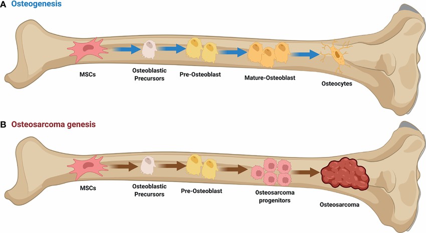 Figure 1. Osteosarcoma. (Xie D, et al.2022)
Osteosarcoma is the most common type of bone malignant tumor, which is developed from mesenchymal cell line. The rapid growth of tumor is due to the direct or indirect formation of tumor bone like tissue and bone tissue in the cartilage stage. Under the influence of external factors (such as viruses), the weight-bearing bones of lower limbs cause cell mutation, which may be related to the formation of osteosarcoma. The typical osteosarcoma originated in the bone. Another completely different type is the osteosarcoma juxtaposed with the bone cortex, which originated from the periosteum and adjacent connective tissue. The latter is rare and has a better prognosis.
The prominent symptom of osteosarcoma is pain at the tumor site, which is caused by tumor tissue erosion and dissolution of bone cortex.
| Symptoms | Descriptions |
| Pain | Pain in different degrees at the tumor site is a very common and obvious symptom of osteosarcoma, which is caused by the destruction of bone cortex by swollen tumor tissue and stimulation of periosteal nerve endings. The pain can develop from intermittent in the early stage to continuous after several weeks, and the degree of pain can be enhanced. Pain avoidance claudication may occur in patients with lower limb pain. |
| Mass | With the development of the disease, local swelling may occur, and a lump may be touched in the painful part of the limb, accompanied by obvious tenderness. If the mass grows rapidly, it can be found from the appearance. The skin temperature on the surface of the mass increases and the superficial veins are exposed. There may be different degrees of tenderness on the surface of the mass and adjacent soft tissues. The hardness of the mass varies with the degree of ossification. The mass increases, resulting in limited joint activity and muscle atrophy. |
| Limping | Pain avoidance claudication caused by limb pain is aggravated with the progress of the disease, and the patients with longer illness time can have limited joint activity and muscle atrophy. |
| General Condition | When the diagnosis is clear, the general condition is generally poor, manifested as fever, discomfort, weight loss, anemia and even failure. In some cases, the tumor grew rapidly, and lung metastasis occurred in the early stage, resulting in the deterioration of the general condition. Pathological fractures at the tumor site make the symptoms more obvious. |
In the case of osteogenic osteosarcoma, the increase of bone derived alkaline phosphatase in the blood can be detected in the early stage, which is related to the osteogenic effect of the tumor. Pathological diagnosis is the basis of treatment. When considering the diagnosis of osteosarcoma, it is of great significance to carry out in vivo tissue examination and get the confirmation of pathological examination as soon as possible.
| Symptoms | Descriptions |
| X-ray photography | The typical x-ray findings of osteosarcoma are characterized by both new bone formation and bone destruction. Most of the tumors are located in the metaphysis of the long tubular bone, with unclear edges, trabecular bone destruction, and increased tumor tissue density. After penetrating the bone cortex, the tumor will push up the periosteum, resulting in the characteristic X-ray signs of the disease -- Codman's sleeved triangle (Codman triangle). This phenomenon can be seen in some patients with osteomyelitis and Ewing sarcoma, and is very typical in osteosarcoma. The shadow of tumor infiltrating soft tissue can be seen in the late stage, and pathological fractures can be seen in some cases. |
| CT scan and MRI examination | It is an effective means to judge the nature and scope of bone tumor and whether there is surrounding soft tissue infiltration. It can early detect the metastatic lesions of lung and other organs. It is a routine item of clinical examination of osteosarcoma. |
| Radionuclide Bone Scan | It is a common method for early detection and late differentiation of metastatic lesions. |
Osteosarcoma is still a disease with high mortality of malignant tumors in children and adolescents, but early detection and timely treatment have greatly improved the survival rate of the disease.
After the pathological diagnosis of osteosarcoma, the early stage of chemotherapy or radiation therapy is started. Resection of tumor tissue is an important step in the treatment of osteosarcoma. With the improvement of tumor surgery technology and the development of implant research, limb preservation therapy has shown a good therapeutic prospect. Consolidation chemotherapy or radiotherapy after tumor tissue resection is very important to control tumor metastasis and improve survival rate.
Radical surgery should be performed in the treatment of osteosarcoma. If possible, extensive local resection can be performed to preserve the limb. In addition, biopsy should be done before amputation.
Immunotherapy is intravenous infusion of lymphocytes or interferon and transfer factor, but the efficacy is uncertain.
The closer the tumor is to the trunk, the higher the mortality. As for the relationship between the type of tumor and the degree of vascular abundance and prognosis, it is difficult to judge. Attention should also be paid to the immune response of patients. Some advanced tumors can survive for a long time without recurrence after radiotherapy, and lung metastasis can dissipate. This may be related to immune response. The key factors affecting the prognosis are early diagnosis, whether the tumor is completely removed, chemotherapy and radiotherapy before and after operation. In addition, it is also related to the type and size of tumor cells, the increase of serum alkaline phosphatase before and after operation, and whether local lymph nodes are involved. Early detection through understanding symptoms and regular physical examination is still essential to improve the chances of successful treatment.
Reference
| Cat. No. | Product Name | Host | Isotype | Application | |
| DMAB4987 | Magic?? Anti-D240 monoclonal antibody, clone E3-41 | Mouse | IgG1 | IHC | Inquiry |
| DMAB5049MH | Magic?? Anti-CDKN1A monoclonal antibody, clone TX119 | Mouse | IgG1 | FC | Inquiry |
| CPBT-38926RM | Anti-LIPG (C-terminal) polyclonal antibody | Rabbit | IgG | IHC-P, WB | Inquiry |
| DPAB-TJ030 | Anti-SLC22A18 polyclonal antibody | Goat | IgG | WB, IF, sELISA | Inquiry |
| DPAB-DC122 | Anti-CTDSP2 (aa 1-99) polyclonal antibody | Mouse | WB, ELISA | Inquiry | |
| DPABH-11075 | Rabbit Anti-Human FOS Polyclonal antibody | Rabbit | IgG | WB, IHC, IF, ELISA | Inquiry |
| DPABH-20692 | Anti-CCSER2 (aa 739-829) polyclonal antibody | Rabbit | IgG | IHC-P | Inquiry |
| Cat. No. | Product Name | Size | Species Reactivity | Application | Detection Method | |
| DEIA-NS2307-133 | Mouse FOS(V-Fos FBJ Murine Osteosarcoma Viral Oncogene Homolog) ELISA Kit | 96T | Mouse | Quantitative | sELISA | Inquiry |
| Cat. No. | Product Name | Size | Target | Species | |
| DAG-P0001 | Human AKAP12 peptide | / | / | / | Inquiry |
| DAG-P1849 | Human OS9 peptide | / | / | / | Inquiry |
Loading ......
