
Inquiry Basket
 Loading ......
In this chapter we describe steps in preparing a quantitative multiplex immunoassay in a micro-plate format based upon the A2 (A-squared) technology. Unlike conventional antibody microarrays in which antibodies are printed directly onto a substrate, the A2 uses oligonucleotide tethering to attach antibodies to the surface. First, a polypropylene 96-well plate is surface treated for covalent attachment of capture oligonucleotides. Amine-terminated capture oligonucleotides are printed in a 7 × 6 array pattern in each well of the plate (A2 Plate).
Next, the complementary oligonucleotides are conjugated to selected antibodies to form the oligo–antibody conjugate.
The conjugates are pooled and added to the plate under hybridization conditions for the creation of the antibody array.
Once prepared, standard immunoassays may be conducted in multiplex.
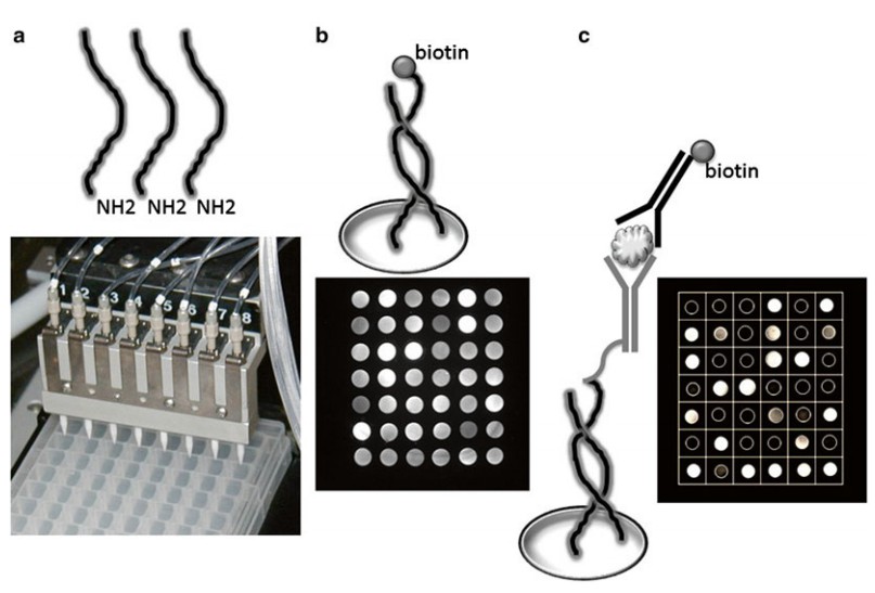 Figure 1. Close-up of the printing process.
What is the advantage of the A2 approach? First, direct immobilization of antibodies often leads to random adsorption onto the surface and as pointed out by Cho et al., “random immobilization results in a low rate (e.g., 5–10 %) of the active antibody density, that is, those that can participate in the binding reaction, which decreases even further when the substance to be analyzed is large in molecular size.”. Thus, stochastic conditions may result in reduced capture efficiency due to steric hindrance or potential blocking of the antibody binding site. Furthermore, printing of proteins can lead to significant variation in spot morphology and protein density thereby reducing array quality. Alternatively, the printing of well characterized capture oligonucleotides offers several advantages. First, oligonucleotide arrays are highly reproducible to produce and extremely stable. Oligonucleotide array plates when properly stored have a shelf-life extending beyond 5 years. Because the formation of duplex DNA is thermodynamically controlled the density of the tethered oligo– antibody conjugates is relatively constant from spot to spot. As a result, the variation of immobilized antibody among spots is reduced which in turn aids in lowering assay imprecision.
The chapter is divided into three main methodologies: oligo-nucleotide–antibody conjugation, hybridization, and performing a multiplex immunoassay.
Materials and Equipment
Micro Centrifuge: 400 × g.
Rotator: end-over-end type or nutating mixer.
Pipettors: single channel.
Pipet Tips: 100–1,000 μL capacity.
Glass pipet: 5 mL.
UV/VIS Spectrophotometer.
Cuvettes.
Micro BCA Protein Assay Kit.
Capture Antibody: 100 μg, purified at a concentration of ≥1 mg/ mL ( see Note 4). Storage buffer must be free of reactive thiols.
Hybridization Buffer A: contains formamide ( see Note 5); avoid contact. Follow proper laboratory safety procedures for handling and storage. Store at 2–8 °C. Hybridization Buffer B: supplied for direct use. Store at 2–8 °C.
Biotinylated Reference Oligo: supplied ready for use. Store at 2–8 °C.
Wash Buffer: tris buffered saline with Tween 20, pH 8.0, 0.05 M Tris –HCl, 0.138 M NaCl, 0.0027 M KCl, Tween 20, 0.05 %. Store at room temperature.
Materials and Equipment
A2 Plate.
Polypropylene Centrifuge Tube, 15 mL, conical.
Platform Shaker (gyratory).
Pipettors: single channel and multichannel.
Pipet Tips: 0–200 μL capacity.
Reagents
Streptavidin Alkaline Phosphatase: dilute 1:20,000 (v/v) in TBST to prepare a working stock solution ( see Note 6).
Streptavidin SureLight P-1: dissolve 150 μg solid with 150 μL distilled water to prepare 50× primary stock concentrate. Dilute stock 1:50 (v/v) in TBST just prior to use as a working stock solution.
Materials and Equipment
A2 Capture Antibody Array Plate: previously prepared.
Platform Shaker (gyratory).
Pipettors: single channel or multichannel.
Pipet tips: 0–200 μL capacity.
A2 MicroArray Reader:
Optical Cube A installed, Ex 545 nm, Em 650 nm and/or Optical Cube B installed, Ex 480 nm, Em 535 nm ( see Note 7).
This protocol is for the preparation of oligonucleotide–antibody conjugates for use with the A2® Plate Array. Approximately 2 OD260 nm units of an activated oligonucleotide are reacted with 100 μg of purified antibody (IgG). The process involves adsorption of the immunoglobulin onto a solid-phase held within a chro matographic column (or large spin-column). Next, initiator is added to modify the antibody with reactive sulfhydryl (-SH) groups. Finally, the SMCC-oligonucleotide is added which couples to the thiolated antibody to form the oligo–antibody conjugate. The resulting conjugate still bound to the solid-support is rinsed free of reactants; and subsequently eluted in purified form from the column ready for use.
Protocol
Solid-Phase: bring to room temperature prior to use; gently mix slurry to dispense.
Spin Column: place bottom cap on column tip and insert capped spin column into a collection tube.
Prepare Spin Columns
Prepare Antibody Binding
Prepare Protein Initiator
Prepare Activated Oligo
Prepare Oligo–Antibody Conjugate
This protocol is for the creation of the A2 Capture Antibody Array by hybridization of the previously prepared Oligonucleotide– Antibody Conjugates. The conjugates are pooled into a hybridization cocktail prepared by mixing Hybridization Buffers A and B; and then applied to the A2 Plate. The number of plates that can be prepared depends upon the conjugate yield and desired loading.
Protocol
Hybridization
This protocol is for the performance of an Enzyme Linked Immunosorbent Assay (ELISA), using A2® Plate with immobilized oligo–antibody array. The previously prepared oligo–antibody conjugates are pooled and applied to the A2 Plate (A21002) under hybridization conditions, resulting in the self-assembly of a capture antibody array in each well of the 96-well plate. After rinsing, the plate is ready to perform the multiplex ELISA. An example of a quantitative multiplex ELISA using this protocol is provided.
Reference
Loading ......
