
Inquiry Basket
 Loading ......
Morphine and other opioids can induce a variety of pharmacological effects. In the central nervous system, they can cause strong analgesia, excitement, sedation, endocrine disorders, respiratory inhibition and so on. In addition, they cause muscle spasms and release histamine in the peripheral nervous system.
The primary metabolic pathway of morphine is glucuronidation via UGT2B7 to form its major metabolite morphine-3-glucuronide and its minor metabolite morphine-6-glucuronide. Morphine can also be converted to normorphine through N-demethylation by CYP3A4 and CYP2C8, which is further metabolized to two glucuronides. Morphine can also be metabolized by SULT1A3 to minor sulfate metabolites. Morphine-6-glucuronide is thought to be active and may partially enhance the analgesic effect of morphine. Some researchers have suggested that its analgesic effect may be small because it accounts for only 10% of the circulating metabolites in plasma and because it does not cross the blood-brain barrier as readily as morphine. However, some studies have shown that the analgesic effect of morphine-6-glucuronide does not depend on its plasma concentration, and that it can be used alone as a drug and is well tolerated. In contrast morphine-3-glucuronide not only shows no analgesic effect, but has also been shown to antagonize the effects of morphine-6-glucuronide. Studies have shown that morphine-3-glucuronide causes neuroleptic effects such as hyperalgesia, allodynia and myoclonus.
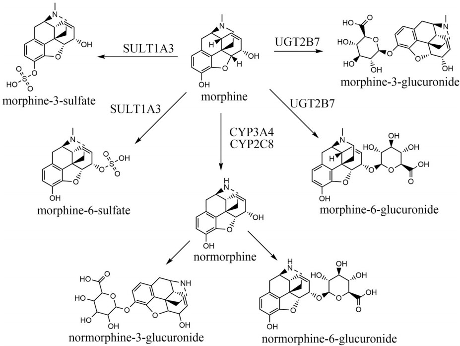 Figure 1. Schematic of morphine metabolism
(Source: Coates S, et al. 2023)
Opioids typically have a narrow therapeutic range, and at normal doses, co-administration of opioids with drugs that may inhibit opioid metabolism may result in adverse drug reactions. Conversely, co-administration of opioids with drugs that may induce opioid metabolism may result in decreased efficacy. Methadone has been shown to interfere with the pharmacokinetics of morphine and reduce the analgesic effect of morphine. Patients on clinical methadone treatment required a higher dose of morphine to achieve the same level of pain relief as patients not on methadone. The formation of morphine-3-glucuronide and morphine-6-glucuronide was reduced 2.4-fold and 3.5-fold, respectively, by co-administration of methadone and codeine, whereas plasma morphine levels were unchanged. The reason for this may be that codeine continues to be metabolized to morphine, whereas methadone inhibits UGT2B7 and UGT2B4, thereby reducing morphine glucuronide levels. Rifampin also has an effect on morphine pharmacokinetics, with rifampin inducing CYP3A4, leading to increased production of normorphine and decreased formation of other metabolites. In the presence of chlorpromazine, morphine AUC increased 2-fold, while in the presence of amitriptyline, morphine AUC increased 1.29-fold. Both drugs caused a significant increase in morphine-induced pain relief in a potentially addictive manner. Both drugs were also found to inhibit UGT2B7 metabolism and to inhibit the formation of morphine-3-glucuronide and morphine-6-glucuronide.
Opioids depress the nervous system by acting on opioid receptors in the brain, spinal cord, and periphery. Upon binding of endogenous or exogenous morphine molecules to opioid receptors, Go or Gi proteins are activated and subsequently phosphorylated by a family of kinases known as G protein-coupled receptor kinases (GRKs). This induces molecular changes within the cell, including binding to β-arrestin. GTP on the α-subunit of the G-protein binds to the opioid receptor, activates it, and dissociates from the βγ-subunit. Both α-GTP and the dimeric βγ subunit are involved in intracellular signaling, inhibit adenylate cyclase activity, decrease intracellular cyclic adenosine monophosphate (cAMP) levels, and inhibit protein kinase A activity. α-GTP also activates the phospholipase C (PLC) and mitogen-activated protein kinase (MAPK) pathways, triggering downstream signaling to increase endoplasmic reticulum calcium release and activate calcium-dependent signaling. It has also been found to activate potassium channels, leading to increased cellular hyperpolarization and indirectly decreasing cellular excitability. βγ dimers directly block calcium channels, thereby decreasing intracellular calcium concentration and inhibiting other neurotransmitters.
Chronic exposure to morphine induces phosphorylation of opioid receptors by GRKs, and this phosphorylation prepares opioid receptors for binding to arrestin. Binding of arrestin blocks further G protein-mediated signaling, leading to opioid receptor desensitization.
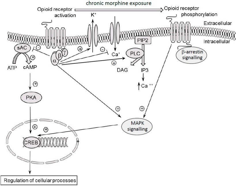 Figure 2. Molecular mechanisms of morphine action
(Source: Listos J, et al. 2019)
Desensitization is a diminished response due to receptor internalization or decreased surface receptor activity after repeated use of morphine or its analogs. Acute doses of morphine reduce cyclic adenosine monophosphate (cAMP) by altering calcium and potassium ion channels, resulting in hyperpolarization. Prolonged use of morphine may lead to adaptive changes observed within opioid receptors, thereby decreasing receptor responsiveness. Opioid receptor trafficking plays a critical role in the development of morphine tolerance, and changes in receptor trafficking are closely linked to the dynamic movement of these receptors to and from the cell surface. Prolonged exposure to morphine alters receptor transport, resulting in a decrease in the number of available surface receptors, which can lead to morphine tolerance. MOR desensitization is also associated with dysregulation of cell membrane proteins (β-arrestin-1 and β-arrestin-2) that bind to opioid receptors and alter their phosphorylation via serine/threonine kinases. Second messenger signaling is inhibited by β-arrestin activation, ultimately leading to receptor desensitization.
Morphine can destroy the intracellular redox balance, but the degree of change is related to age, sex, dose, medication time and other drug interactions. Morphine can induce the formation of free radicals and reduce the ability of antioxidant defense, resulting in cell damage. Nitric oxide is produced by L-arginine under the action of nitric oxide synthase (NOS), which can be divided into inducible NOS (iNOS), endothelial NOS (eNOS), and neuronal NOS (nNOS). It acts as a neurotransmitter in the signal pathway between neurons, glial cells and cerebral vessels.
Peroxynitrite (PN) is involved in pain modulation, opioid-induced analgesia, and antinociceptive tolerance through the formation of superoxide and nitric oxide precursors involving NOS, NADPH oxidase, and spinal manganese superoxide dismutase (MnSOD). Activation of NOS and NADPH-oxidase leads to NO and superoxide production, respectively, whereas inactivation of MnSOD leads to PN formation, and inhibition of NOS and MnSOD activity has been shown to prevent morphine-induced antinociceptive tolerance. Morphine causes a decrease in glutathione (GSH) levels in the human brain, and long-term use interferes with the activity of catalase (CAT), superoxide dismutase (SOD), and glutathione peroxidase (GSHPx). Morphine was also found to induce increased expression of thioredoxin-1 (Trx-1), a redox-regulated protein and a neurotrophic cofactor that inhibits tolerance to the analgesic effects of morphine.
Opioid tolerance and dependence are a biphasic pattern divided into an induction phase and a manifestation phase. It has been found that the antinociceptive of NOS deficient mice is decreased, and the use of NOS inhibitors in morphine dependent mice can also reduce naloxone-induced withdrawal jump and diarrhea. In addition, the researchers also found that the abnormal excitation of neurons or synapses in the central nervous system caused by the activation of NO-cGMP pathway is associated with chronic pain. Interestingly, L-arginine can mimic chronic pain by activating the NO-cGMP pathway. NO-cGMP signaling has also been implicated in reward memory. Thus, NOS inhibitors may suppress morphine-dependent expression by inhibiting the NO-cyclic GMP system, which is thought to mediate the effects of excitatory amino acid receptor activation.
The involvement of N-methyl-D-aspartate (NMDA) receptors in neuronal development, memory formation, learning, and hyperactivity or dysfunction of NMDARs can lead to disease pathology. These receptors have been implicated in a variety of nervous system disorders, including ischemic brain injury, neurodegenerative disease, depression, schizophrenia and pain. In a mouse model, the chronic use of morphine increases the activity of the NMDA receptor in the mouse brain. Repeated and prolonged exposure to opioids may result in altered neuronal plasticity, but the use of NMDA antagonists may affect dependence and tolerance to opioids and attenuate or block behavioral changes associated with morphine withdrawal following naloxone administration.
In the central nervous system, PPAR is mainly related to lipid metabolism, nerve cell differentiation, cell death, inflammation and nerve degeneration. PPAR contains three isozymes (α, β and γ). PPARα and PPARγ are widely expressed in the lateral hypothalamic area, which is located in the ventral tegmental area (VTA) and controls the release of dopamine from the VTA to the Nucleus accumbens (NAc). These projections are associated with reward pathways and have a potential role in addiction. PPARγ is a ligand-activated transcription factor that regulates genes required for cellular differentiation and various metabolic processes. Activation of macrophages by PPARγ agonists results in cellular morphological changes and inhibition of nitric oxide, and also induces apoptosis by interfering with the NF-κB pathway. PPARγ agonists have been used in animal models of drug addiction, and thiazolidinediones reduce alcohol consumption and withdrawal symptoms in animals. PPARγ has been shown to play a key role in morphine tolerance and dependence, and pioglitazone improves symptoms of morphine tolerance and dependence in rats, whereas the effects of pioglitazone are attenuated or even reversed using PPARγ antagonists.
Prolonged use of morphine induces a neuroinflammatory response by activating glial cells and releasing pro-inflammatory cytokines. These inflammatory mediators alter the excitability and plasticity of neurons, reduce the efficacy of morphine and lead to tolerance. Morphine binds to Toll-like receptor 4 (TLR4), triggering the release of proinflammatory cytokines and glutamate, thereby increasing morphine tolerance. The neuroprotective effect of morphine involves the activation of PI3K pathway, which promotes cell survival and inhibits apoptosis through downstream signal target Akt, while Akt activates NF-κB through phosphorylation of IkB kinase, thus promoting cell survival. Opioids also induce tumor necrosis factor-α (TNF-α), which in turn activates macrophages, leading to cell proliferation, extracellular matrix remodeling, and cytokine release, thereby activating the MAPK signaling cascade. Morphine increases TNF-α expression in astrocytes, microglia and U937.
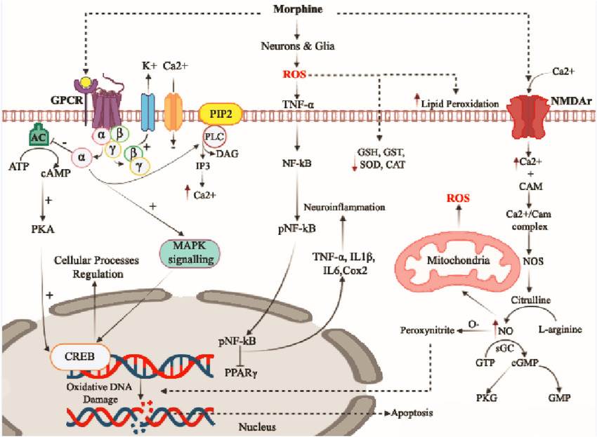 Figure 3. A schematic diagram showing multiple downstream signaling pathways that mediate morphine-induced tolerance and dependence
(Source: Badshah I, et al. 2023)
The environment in which the addictive substance is used can have a significant impact on the individual; when the individual is exposed to some drug-related stimuli, it can cause cravings for the drug, and the increased rewarding effect of the addictive substance can also lead to relapse. Psychological addiction can be assessed by examining behavioral sensitization, which refers to the exacerbation of behavioral or motor responses after repeated exposure to certain drugs of abuse in drug-free environments, and its progression involves neuroadaptive changes, including dysregulation of dopaminergic and glutamatergic neurotransmission. Much of the attention in drug abuse has focused on the midbrain-limbic dopamine pathway, which includes dopaminergic neurons located in the midbrain VTA and their projections to the limbic forebrain and NAc. Morphine indirectly mediates dopamine efflux in the nucleus accumbens by binding to MORs. The sensitizing effect of morphine is associated with increased dopamine release in mesolimbic regions with dopaminergic D1 receptors.
References
| Target | Cat. No. | Product Name | Size | Species Reactivity | Application | Detection Sample | |
| Morphine | DEIA2086NS | Morphine ELISA Kit | 96T | N/A | Quantitative | Plasma, serum, and urine | Inquiry |
| DEIA04876 | Canine Morphine Specific Direct ELISA Kit | 96T | Dog | Quantitative | Whole blood oral fluids, serum, plasma, urine | Inquiry | |
| DEIA2085 | Morphine ( serum/blood ) ELISA Kit | 96T | Quantitative | Serum, whole blood | Inquiry | ||
| DEIA2086 | Morphine ELISA Kit | 96T | Quantitative | Whole blood, oral fluids, serum, plasma and urine | Inquiry |
| Target | Cat. No. | Product Name | Expression System | Tag/Conjugate | Application | |
| Morphine | DAG1243 | Morphine [HRP] | N/A | HRP | N/A | Inquiry |
| DAGA-303B | Morphine [BSA] | N/A | BSA | LFIA | Inquiry | |
| DAGF-147 | Morphine (MS Contin) [BSA] | Synthetic | BSA | ELISA, LF | Inquiry | |
| DAGF-148 | Morphine (MS Contin) Hapten/Protein Ratio=40:1 [BSA] | Synthetic | BSA 40:1 | ELISA, LF | Inquiry | |
| DAGF-171 | Morphine (MS Contin) Hapten/Protein Ratio=70:1 [BSA] | Synthetic | BSA | ELISA, LF | Inquiry |
Loading ......
