


Inquiry Basket
 Loading ......
HSV-1 and HSV-2 are common human pathogens. The global seroprevalence of HSV-1 is about 67% while HSV-2 is 13%. HSV-1 and HSV-2 infection are both acquired via close contact and result in a life-long infection. HSV-1 is usually acquired earlier in life through orolabial mucosa, whereas HSV-2 is usually acquired later in life by sexual transmission. Infection with HSV-1 usually confers immunity to HSV-1 reinfection, but not HSV-2. HSV-1 also predisposes an individual to human immunodeficiency syndrome (AIDS).
Creative Diagnostics provides high-quality antibodies, antigens, and various reagents related to HSV to support your research and development needs.
Latency establishment and its periodic reactivation are hallmark characteristics of HSV infection. Replication at the site of entry is followed by transport of intact virus particles (or nucleocapsids) by retrograde axonal transport to sensory (HSV-1 predominantly in the trigeminal ganglia, HSV-2 in the sacral dorsal root ganglia) or autonomic ganglia, where the virus persists in a latent state. After reactivation, it is carried by anterograde transport to the site of the original infection, resulting in a recurrent episode of symptoms. The reactivation is usually caused by local trauma to the infected nerve or by a systemic factor such as stress, fever, ultraviolet light, menstruation or hormonal changes.
HSV infects epithelial cells of the mucosa or skin during primary infection and establishes latency mostly in neurons of the PNS. HSV is spread by close contact. The virus usually infects the mucosa or epidermis after minor breaches of the skin surface. Following efficient replication in epithelial cells, HSV is transported to the peripheral nerve endings, and taken up by axonal transport to the neuronal cell bodies in a retrograde fashion.
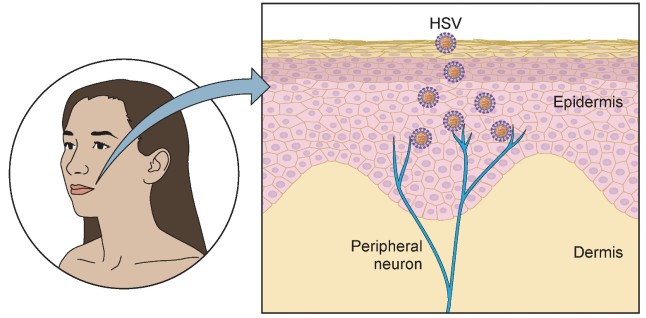 Figure 1. Initial steps of HSV primary infection.
(Source: Zhu S, et al. 2021)
Lytic Replication
Latent Phase
Gene expression during the latent and lytic cycles of HSV is not mutually exclusive; instead, there are intermediate stages with varying levels of viral gene expression. HSV may undergo lytic transcription in some neurons, which can be blocked by the host or lead to the production of infectious virions, while remaining latent in others.
VP16 plays a central role in initiating lytic viral gene expression. After the viral envelope fuses with the cell membrane, VP16 and other tegument proteins enter the nucleus separately. However, due to the long distance from the neurite end to the neuronal cell body, they may not reach the neuronal nucleus promptly, resulting in low levels of VP16. This limits effective expression of immediate early (IE) genes and promotes entry into latency.
Epigenetic modifications play a crucial role in the molecular regulation of latency establishment, maintenance, and reactivation. The incoming HSV genome lacks histones and methylation. Once the viral DNA enters the cell and is detected by pattern recognition receptors (PRRs), various enzymes deposit histones and histone modifications on the HSV genome, restricting gene expression.
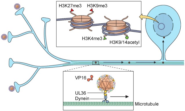 Figure 2. Establishment of HSV latency in neurons.
(Source: Zhu S, et al. 2021)
How viral genes become reactivated and begin to be expressed from a silenced genome may be linked to the neuronal stress response and the JNK pathway. The VP16 promoter contains Egr-1/Sp1 binding sites that respond to transcription factors expressed during neuronal stress. This likely helps promote VP16 expression under stress conditions, triggering viral reactivation in neurons. Various stimuli causing neuronal stress have been shown in animal models and cell cultures to induce reactivation, including removal or inhibition of neurotrophic factors, exposure to UV light, inhibition of phosphoinositide 3-kinase (PI3K), inhibition of histone deacetylases, and activation of JNK. In humans, UV light exposure, hormone level changes, and fever can trigger HSV reactivation. One cytokine expressed under these conditions, IL-1β, has been shown to induce HSV-1 reactivation in latently infected mouse neurons via a dual-leucine-zipper kinase (DLK)-dependent pathway. The mechanism by which JNK induces gene expression from a repressed genome appears to involve phosphorylation of serine residues in histones, allowing gene expression even in the presence of repressive methylation on histone lysines—a process called the methyl-phospho switch.
 Figure 3. Mechanisms of regulation of viral genes during latency and reactivation in neurons.
(Source: Fu H, et al. 2025)
Animation
Studies in mouse neurons suggest that reactivation occurs in two phases.
The first phase involves low-level genome-wide expression without viral protein production, leading to protein translation.
Productive Reactivation
VP16 expression is required for the second phase, which induces IE gene expression and triggers a gene expression cascade similar to that seen during lytic infection.
After reactivation, new infectious viral particles are produced and travel anterogradely to the skin or mucosa, causing the typical herpes lesions.
Involvement of the oral cavity usually happens at the time of primary infection with HSV-1. Symptomatic cases can be as varied as fever, ulcers on the throat, blisters and pain in the mouth and gums. Gingivostomatitis is very common in children and adolescents who acquire HSV-1. The lips, tongue, gums, buccal mucosa and palate are often involved when patients are symptomatic. Genital herpes is caused in most cases by HSV-2. The ulcerative blisters form on the penis, cervix, vulva, vagina or perineum. Oral and genital recurrences are usually preceded by a prodromal stage, in which the patient experiences pain, burning, itching or tingling.
References
Loading ......
