
Inquiry Basket
 Loading ......
Exosomes are extracellular vesicles (EVs) produced by endosomes in eukaryotic cells, which have attracted much attention in life science research and biotechnology because of their involvement in intercellular communication in a variety of normal and pathological functions.
The double invagination of the plasma membrane together with the luminal vesicles (ILVs) forms intracellular multivesicular vesicles (MVBs), which are expelled from the cell after a series of material sorting, which is the process of exosome formation. ILVs can carry lipids, proteins, and nucleic acids from the parent cell. Their formation is driven by the endosomal sorting complex required for trafficking (ESCRT) and the associated proteins ALG2-interacting protein X (ALIX) and tumor susceptibility gene 101 (TSG 101), which ubiquitinate MVB membrane proteins and induce inward budding of MVB membranes. In addition, ILV can be produced in an ESCRT-independent manner. Ceramide lipids form lipid raft microdomains that induce MVB membrane outgrowth. Researchers have also implicated the involvement of the Ras-related proteins GTPase Rab, tetraspanins, syndecan-1, and phospholipids in exosome biogenesis.
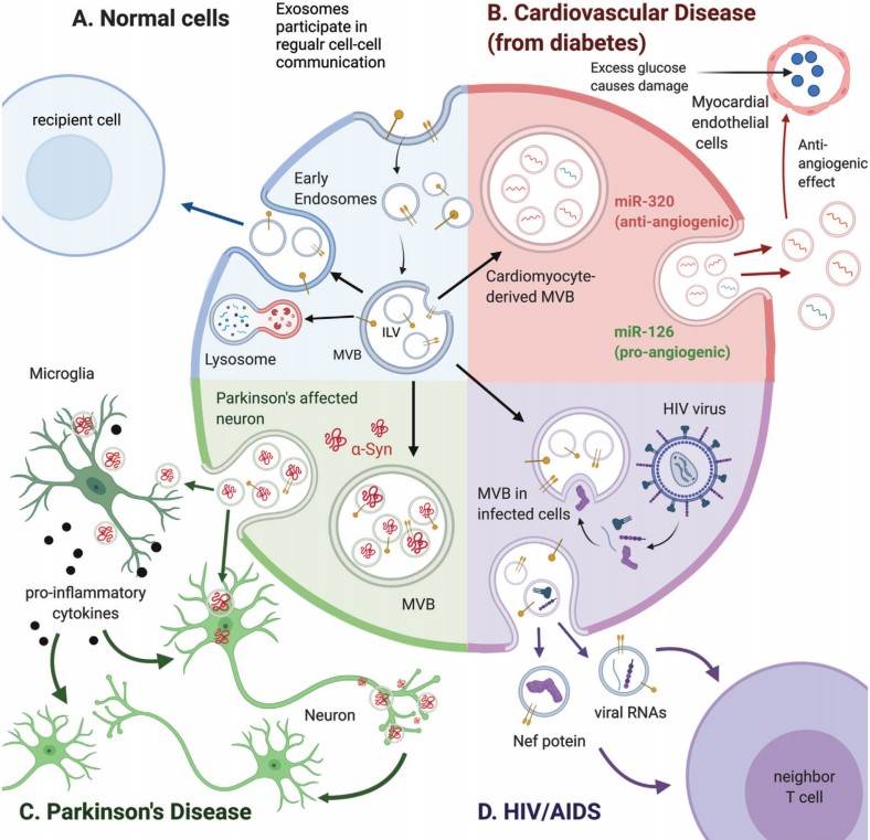 Figure 1. Exosome biogenesis
(Source: Lai JJ, et al. 2022)
Because of the heterogeneity between and within exosomes types, and the feature overlap with other EVs, it is difficult to distinguish exosomes from other EVs. Different exosomes separation techniques have been developed based on the physicochemical properties of exosomes as well as carrying surface biomarkers. Non-specific separation based on the physical properties of exosomes includes ultracentrifugation, size-exclusion chromatography (SEC), and polymer precipitation, and specific enrichment based on the biological properties of exosomes includes immunoaffinity.
Ultracentrifugation has been widely used to isolate exosomes from a variety of sources (e.g., cell culture media, serum, saliva, urine, and cerebrospinal fluid) and is considered to be the gold standard for the isolation of exosomes; however, there are limitations to the method, such as cumbersome and costly instrumentation, long and laborious processing, contamination of aggregated proteins and ribonucleoprotein particles, and the need for a large number of samples.
Polymers precipitate by reducing the hydration of exosomes to reduce the solubility and thus cause precipitation, the precipitated products can be easily separated by low centrifugal force. This method has a short extraction time and can meet the needs of different conditions. However, this method is expensive for large-scale use, lack of specificity and low purity. Heterogeneous polymer particles tend to be produced in the process of co-precipitation, high likelihood of remaining extracellular contaminants due to reagents that similarly reduce the solubility of vesicles and proteins, also co-precipitate a variety of water-soluble substances such as nucleic acids, lipoproteins, proteins, and even viruses.
Size-exclusion chromatography (SEC) separates vesicles and other molecules based on their size by gel filtration, where small molecules diffuse into the pores as the sample enters the gel, while larger molecules elute directly. Therefore, the larger molecules leave the column earlier than the small ones, which means that the residence time of a molecule is correlated with its size. The size-exclusion column contains resins with a pore size of approximately 75 nm, allowing larger vesicles (>75 nm) to quickly pass through and elute out of the voids, while proteins and other smaller molecules temporarily diffuse into the pores and cannot pass through. SEC has little damage to exosomes and low demand for sample volume, but the exosomes prepared by SEC column usually show wider size distribution, especially in a smaller size range, which indicates the existence of pollutants with similar size to exosomes, such as protein aggregates and lipoproteins.
Immunoaffinity targets marker proteins on the surface of the exosome membrane and utilizes specific antibodies to capture common markers of exosomes. In theory, any protein highly present on the exosome membrane and the lack of corresponding free components in the extracellular fluid can capture the exosomes based on the principle of immune affinity.
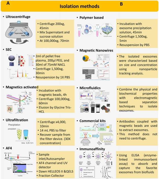 Figure 2. Different techniques used for isolation of exosomes
(Source: Mahgoub EO, et al. 2020)
Exosomes is helpful in the diagnosis of diseases, they are present in all biological fluids, and the components of complex goods containing exosome can be easily obtained by liquid biopsy, which is potentially valuable in diagnosing patients with cancer and other diseases, as well as in determining prognosis. The disease progression and therapeutic effect can also be determined by multicomponent analysis of exosomes. For example, in cancer research, researchers have found that there were significant differences in exosomes carrying information between cancer patients and normal healthy people, and have screened for various types of cancer-specific exosomes markers, which can be used as effective biomarkers for staging tumors, identifying molecular mutations, and monitoring the effectiveness of treatments.
Exosomes diagnosis is mainly used in cardiovascular diseases, central nervous system diseases, cancer, follow-up diagnosis applications gradually related to liver, kidney, lung-related diseases. Specific miRNAs carried by exosomes have the potential to assist in the diagnosis of cancer, and have high diagnostic value because exosomes secreted by cancer cells carry carcinogenic or tumor suppressor miRNAs that are significantly different from those of normal cells. For example, the increase of miR21 in circulating exosomes is associated with glioblastoma, pancreatic cancer, colon cancer, colon cancer, liver cancer, breast cancer, ovarian cancer, and esophageal cancer, while the increase of exosomes miR21 in urine is associated with bladder cancer and prostate cancer.
In addition, the surface and intracellular proteins carried by exosomes can also be used as diagnostic markers. It is reported that glypican-1 (GPC1) is rich in exosomes secreted by cancer cells and is related to the diagnosis of pancreatic cancer, breast cancer and colon cancer.
The possibility of combining protein, nucleic acid, and lipid expression levels in exosomes to assess disease progression is being considered, with the potential to improve the specificity and sensitivity of exosomes-based diagnostics using a combination of markers that reflect different aspects of the disease-associated exosome.
The exosomes themselves can be used as drug-carrying vehicles to achieve therapeutic effects. Compared with liposomes, injection of exosome can effectively enter the cells and deliver functional cargo with a minimum immune clearance rate after exogenous drugs are injected into mice. The exosomes are also well tolerated, with no significant side effects from repeated injections. On this basis, researchers have developed engineered exosomes for central nervous system and cancer, which can carry designated miRNA or small interference RNA. These RNAs can effectively bind to target mRNAs and inhibit gene expression in receptor cells, thus achieving therapeutic goals. The RNA in exosomes can be protected from the degradation of ribonuclease in the blood, and the therapeutic exosomes has better systemic retention than liposome and can function at a distance. Studies have shown that there is CD47 on the surface of exosomes, which sends a "don't eat me" signal, protects exosomes from being swallowed, and reduces clearance in blood circulation.
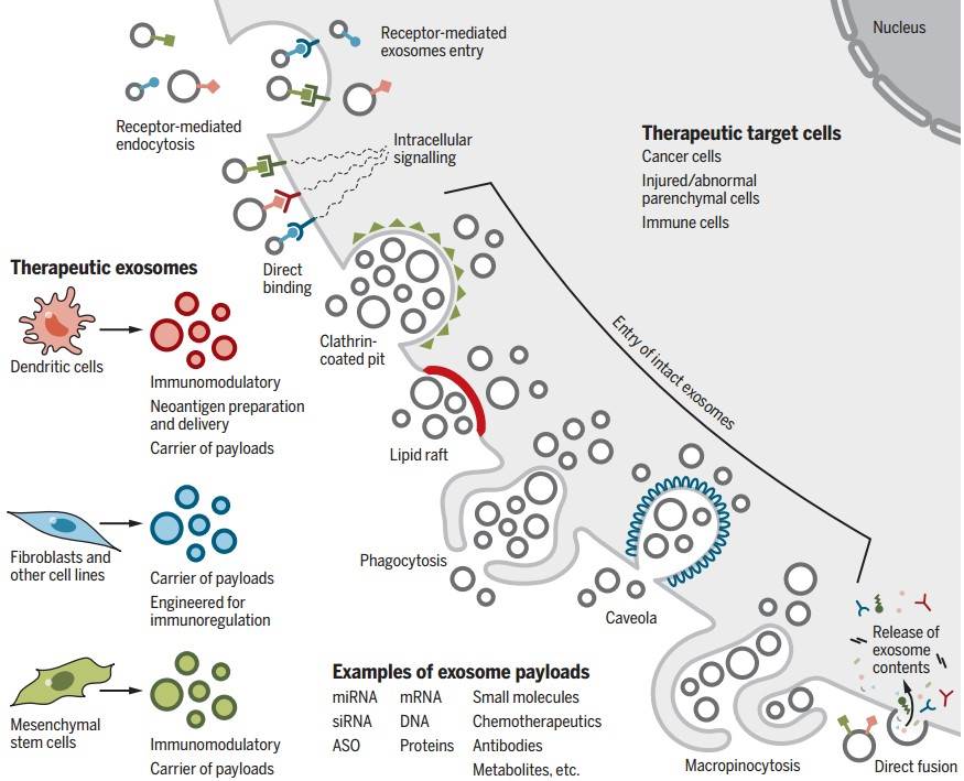 Figure 3. Cellular uptake of therapeutic exosomes
(Source: Kalluri R, et al. 2020)
At present, the biggest challenge of exosomes-based therapy is to solve the heterogeneity of secreted exosomes. Exosomes-based therapy requires a better understanding of its biogenesis, composition, and heterogeneity. Although exosomes extracted from similar cells is considered to have the same composition, the results show that these exosomes may have different molecular composition and targeting moiety. The heterogeneity of exosomes brings additional complexity to exosome design, dose standardization and delivery in clinical applications. Therefore, an in-depth study of the heterogeneity of exosomes is crucial, not only to identify suitable subpopulations for specific therapeutic purposes, but also to prevent side effects associated with heterogeneity. Different isolation methods have an effect on the purity and physicochemical properties of exosomes, so optimizing the isolation method can better preserve the biological activity of exosomes and reduce the associated side effects.
The composition and carrying information of exosomes are closely related to that of the parental cells, so when using exosomes for treatment, we can choose exosomes that are more favorable for treatment based on the characteristics of exosomes isolated from different types of cells. Transplanted myeloid dendritic cells, melanoma and muscle cell-derived exosomes have been shown to have different distribution patterns in the spleen, lung, and liver. In addition, neutrophil-derived exosomes can cross the blood-brain barrier and can be used for drug delivery to the brain and targeting of gliomas.
References
| Target | Cat. No. | Product Name | Expression System | Tag/Conjugate | Application | |
| Exosome | DAG-WT1158 | Purified exosomes from MSC | MSC from Adipose tissue | N/A | Calibration, Control, Flow cytometry, EM | Inquiry |
| DAGA-987 | Purified exosomes from plasma of healthy donors | Plasma of healthy donors | Unconjugated | Calibration, Control, Flow cytometry, EM | Inquiry | |
| DAGA-988 | Purified exosomes from urine of healthy donors | Urine of healthy donors | Unconjugated | Calibration, Control, Flow cytometry, EM | Inquiry | |
| DAGA-989 | Purified exosomes from COLO1 cell culture supernatant | Human Colon Carcinoma cell line COLO1 | Unconjugated | Calibration, Control, Flow cytometry, EM | Inquiry | |
| DAGA-990 | Purified exosomes from MM1 cell culture supernatant | Human Melanoma cell line MM1 | Unconjugated | Calibration, Control, Flow cytometry, EM | Inquiry | |
| DAGA-991 | Purified exosomes from BLCL cell culture supernatant | BLCL21 | Unconjugated | Calibration, Control, Flow cytometry, EM | Inquiry | |
| DAGA-992 | Purified exosomes from HCT116 cell culture supernatant | Human Colon Carcinoma cell line HCT116 | Unconjugated | Calibration, Control, Flow cytometry, EM | Inquiry | |
| DAGA-993 | Purified exosomes from U87 MG cell culture supernatant | Human Glioblastoma-Astrocytoma cell line U87 MG | Unconjugated | Calibration, Control, Flow cytometry, EM | Inquiry | |
| DAGA-994 | Purified exosomes from SK-N-SH cell culture supernatant | Human Neuroblastoma cell line SK-N-SH | Unconjugated | Calibration, Control, Flow cytometry, EM | Inquiry | |
| DAGA-995 | Purified exosomes from PC3 cell culture supernatant | Human Prostate adenocarcinoma grade IV cell line PC3 | Unconjugated | Calibration, Control, Flow cytometry, EM | Inquiry | |
| DAGA-996 | Purified exosomes from BPH-1 cell culture supernatant | Human being prostatic hyperplasia cell line BPH-1 | Unconjugated | Calibration, Control, Flow cytometry, EM | Inquiry | |
| DAGA-997 | Purified exosomes from DAUDI cell culture supernatant | Human Burkitt Lymphoma cell line DAUDI | Unconjugated | Calibration, Control, Flow cytometry, EM | Inquiry | |
| DAGA-998 | Purified exosomes from A549 cell culture supernatant | Lung carcinoma cell line A549 | Unconjugated | Calibration, Control, Flow cytometry, EM | Inquiry | |
| DAGA-999 | Purified exosomes from K562 cell culture supernatant | Pleural effusion, leukemia chronic myelogenous cell line K562 | Unconjugated | Calibration, Control, Flow cytometry, EM | Inquiry | |
| DAGA-1000 | Purified exosomes from B16F10 mouse cell culture supernatant | Mouse melanoma cell line B16F10 | Unconjugated | Calibration, Control, Flow cytometry, EM | Inquiry | |
| DAGA-1001 | Purified exosomes from serum of healthy donors | Serum of healthy donors | Unconjugated | Calibration, Control, Flow cytometry, EM | Inquiry | |
| DAGA-1002 | Purified exosomes from saliva of healthy donors | Saliva of healthy donors | Unconjugated | Calibration, Control, Flow cytometry, EM | Inquiry | |
| DAGA-1003 | Fluorescent labeled Exosome Standards | N/A | FITC | Fluorescence microscopy, Flow cytometry, EM | Inquiry |
Loading ......
