
Inquiry Basket
 Loading ......
Interleukins (IL) are produced by lymphocytes and monocytes and bind to specific receptors on other inflammatory cells and the immune system (B-cells, T-cells, mast cells) to produce a transient response. Macrophages and endothelial cells synthesize interleukins along with tumor necrosis factor (TNF), which also gives the two cytokines the same functional properties and similar behavior. After bacterial invasion, macrophages recognize the outsiders, are activated and secrete IL-1 and TNF. These hormones alter the lining of blood vessels, activate T cells, and circulate to the brain. IL-1 and TNF in the brain promote the synthesis of prostaglandin E2, which alters our internal regulation of body temperature.
CD4+ Th cells can be categorized into different subpopulations based on different cytokine profiles, and the cytokine expression profiles depend on the adjuvanticity of the molecules presented with the antigen and the status of the T cells, as well as the type of antigen-presenting cells (APCs) and cytokines in the microenvironment. Different interleukins can interact with specific Th cells to promote different types of inflammatory responses. For example, effector Th2 cells produce IL-4, IL-5, IL-9, and IL-13 in allergic diseases, and their production of IL-25, IL-31, and IL-33 promotes TH2 responses and inflammation. TH17 cells express IL-6, IL-8, IL-22, and IL-26.
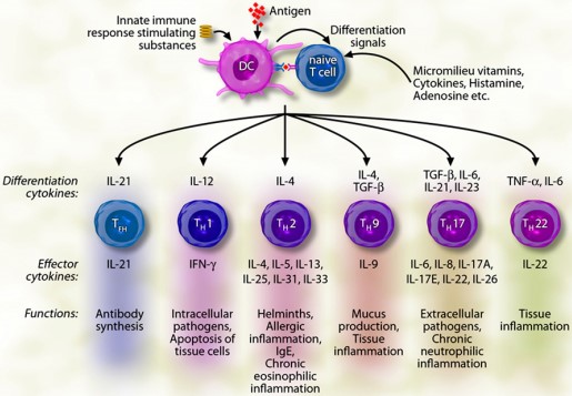 Figure 1. Antigen presentation by DCs to naive T cells induces the T cells to produce ILs and differentiate into TH1, TH2, TH9, TH17, TH22, or follicular TH (TFH) cells
(Source: Akdis M, et al. 2011)
A number of different interleukins have been identified with varying activities and roles in target cells.
Table 1. Some known interleukins and their functions
| Cytokine | Source | Target cell | Major functions |
| IL-1α, IL-1β | Macrophages, monocytes, lymphocytes, keratinocytes, microglia, megakaryocytes, neutrophils, fibroblasts, synovial lining cells | T cells, fibroblasts, epithelial and endothelial cells | Induction of proinflammatory proteins, hematopoiesis, differentiation of TH17 cells |
| IL-2 | CD4+ and CD8+ activated T cells, DCs, NK cells, NKT cells | CD4+ and CD8+ T cells, NK and B cells | Proliferation of effector T and B cells, development of Treg cells, differentiation and proliferation of NK cells and growth factor for B cells |
| IL-3 | T cells, macrophages, NK cells, mast cells, eosinophils, stromal cells | Erythroid progenitors, granulocyte-macrophages progenitors, CD34+ progenitor cells, basophils, eosinophils | Hematopoietic growth factor, activation of basophils and eosinophils |
| IL-4 | TH2 cells, basophils, eosinophils, mast cells, NKT cells, γ/δ T cells. | T and B cells | Induction of TH2 differentiation, IgE class switch, upregulation of class II MHC expression on B cells, upregulation of CD23 and IL-4R, survival factor for B and T cells, role in tissue adhesion and inflammation |
| IL-6 | Endothelial cells, fibroblasts, monocytes/macrophages | Hepatocytes, leukocytes, T cells, B cells, hemopoietic cells | Liver: synthesis of acute phase proteins; leukocytes: trafficking, activation; T cell: differentiation, activation, survival; B cell: differentiation, production of IgG, IgM, IgA hematopoiesis |
| IL-8 | Monocytes, macrophages, neutrophils, lymphocytes, endothelial cells, epithelial cells, fibroblasts, keratinocytes, chondrocytes, synovial cells, hepatocytes | Neutrophils, NK cells, T cells, basophils, eosinophils, endothelial cells | Chemoattractant for neutrophils, NK cells, T cells, basophils, eosinophils; mobilization of hematopoietic stem cells; angiogenesis |
| IL-10 | T cells, B cells, monocytes, macrophages, DCs | Macrophages, monocytes, T cells, B cells, NK cells, mast cells, DC and granulocytes | Immune suppression |
(Source: Akdis M, et al. 2011)
Interleukin 1
IL-1, originally known as the human leukocyte pyrogen, consists of IL-1α and IL-1β, which share similar, albeit minimal, sequence homology but have similar biological properties, although they differ fundamentally in their localization, maturation, and secretion. IL-1α is translated into a biologically active form, whereas IL-1β is translated into pro-IL-1β, which is biologically inactive until processed by caspase-1. IL-1α and IL-1β exert similar effects by binding to the IL-1 type I receptor (IL-1RI).
IL-1β is a circulating factor whose expression is tightly regulated. The main source of IL-1β is myeloid cells, which express the cytokine as a cytoplasmic 31kDa precursor protein (pro-IL-1β) that needs to be converted to the mature form to function. IL-1β is usually activated by caspase-1 cleavage of the N-terminal precursor region of pro-IL-1β. Different lysis sites for neutrophil protease and mast cell chymotrypsin are present in the N-terminal region of pro-IL-1β, and thus these proteases can independently produce mature and active IL-1β. Due to the absence of a pilot sequence, mature IL-1β is released in the environment through an unconventional secretory mechanism independent of the endoplasmic reticulum and Golgi network.
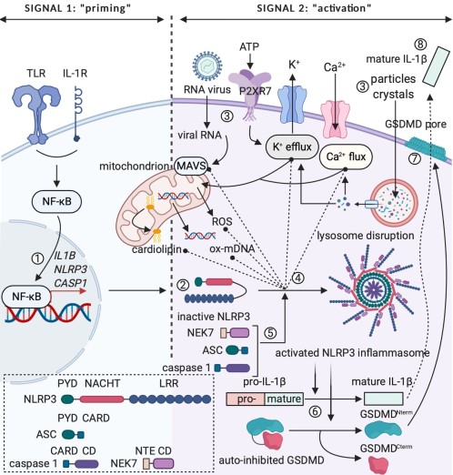 Figure 2. The two-step paradigm of controlled IL-1β release
(Source: Van Den Eeckhout B, et al. 2021)
IL-1 is a potent pro-inflammatory cytokine with multiple potentiating effects on cell proliferation, differentiation and function of many innate and specific immune cells. IL-1 mediates many inflammatory diseases by initiating and enhancing immune and inflammatory responses. There are currently 21 members of the IL-1 superfamily of ligands and receptors, including 11 soluble factors and 10 receptor molecules. Of these soluble factors, seven are pro-inflammatory and four are anti-inflammatory. Two of these anti-inflammatory mediators are receptor agonists and two are receptor antagonists. The IL-1 receptor superfamily is divided into several subgroups: ligand-binding receptor proteins, accessory chains, molecules that inhibit signaling, and orphan receptors.
Interleukin 18
IL-18, a member of the IL-1 family, is expressed by macrophages, Kupffer cells, keratinocytes, dendritic cells and a series of cells. It shares the same structural features as IL-1 and also requires caspase-1 lysis to become a biologically active molecule. IL-18 binds to IL-12 to induce T cells to produce high levels of IFN-γ, whose biological activity can be neutralized by IL-18-binding proteins. IL-18 expression has been associated with rheumatoid arthritis and Crohn's disease activity.
A number of interleukins bind to common γ chain (γc) receptors and are therefore categorized into the γc family, including interleukins 2, 4, 7, 9, 15, and 21. They act mainly as growth and proliferation factors for progenitor and mature cells, but also play a role in lineage-specific cell differentiation.
Interleukin 2
IL-2 is produced primarily by CD4+ T lymphocytes (naïve, memory, and T helper 1) following antigenic stimulation, by type 2 and type 3 innate lymphocytes in the small intestine, and to a lesser extent by activated CD8+ T cells, B cells, and other innate immune entities.
In naïve T lymphocytes, binding of the TCR and co-stimulatory molecules within the immune synapse activates activator protein 1 (AP-1), NFκB, and NFAT. These transcription factors cooperate with constitutive factors to promote IL-2 gene expression. IL-2 transcription occurs within 30 minutes of stimulation and falls to background levels within 24-48 hours. In addition, post-transcriptional regulatory mechanisms further limit the availability of IL-2 mRNA, the levels of which typically peak 4-8 hours after stimulation. The transversion of IL-2 mRNA is mainly controlled by proteins interacting with its AU-rich cis-element (ARE) in the 3'-untranslated region. Nuclear factor 90 (NF90) is activated by protein kinase B (PKB) in response to CD28 co-stimulation or by PKC in response to PMA re-stimulation, and then exported from the nucleus to the cytoplasm. In the cytoplasm, NF90 binds to the ARE and stabilizes IL-2 mRNA, thereby allowing it to be translated.
IL-2 is essential for Treg cell development. IL-2 also acts as a B-cell growth factor, stimulates antibody synthesis, and promotes the proliferation and differentiation of NK cells, thereby enhancing their cytolytic function. Recombinant Human IL-2 Could Be Used for Immunotherapy to Treat Cancer and AIDS.
 Figure 3. IL-2 transcription following stimulation of the TCR and CD28 signaling cascades
(Source: Pol JG, et al. 2020)
Interleukin 4
IL-4 is a monomer produced by Th2 cells, basophils, mast cells and eosinophils. There are two types of IL-4 receptors (IL-4R), type I IL-4R binds only IL-4 and type II IL-4R binds IL-4 and IL-13. IL-4 regulates allergic reactions as well as protective immune responses against parasites. It also inhibits the development of Th1 cells, induces IgE sorting transitions in B cells, and increases the expression of MHC II molecules. In addition, IL-4 upregulates B cell receptors, increases CD23 expression, prolongs the lifespan of T and B cells in culture, and mediates tissue adhesion and inflammation.
Interleukin 8
IL-8 was identified as a neutrophil-specific chemokine and later classified as a member of the CXC chemokine family. A variety of cells can produce IL-8 upon stimulation with IL-1-α, IL-1-β, IL-17, TNF-α, or TLRs, including monocytes, macrophages, neutrophils, lymphocytes, endothelial cells, and epithelial cells. IL-8 production is dependent on upstream NF-κB signaling, while chemokines themselves trigger downstream activation of PI3K and MAPK signaling cascades. The primary function of IL-8 is to bind to two G protein-coupled receptors, CXCR1 and CXCR2, and is responsible for the activation and recruitment of neutrophils to sites of infection or injury. In addition to neutrophils, IL-8 attracts NK cells, T cells, basophils and GM-CSF-catalyzed or IL-3-catalyzed eosinophils. IL-8 concentrations are elevated at sites of inflammation in patients with rheumatoid arthritis, respiratory syncytial virus infection, or chronic obstructive pulmonary disease (COPD).
In the tumor context, IL-8 has a dual pro-tumorigenic effect, on the one hand directly affecting the tumor cells themselves and on the other hand altering the composition of the tumor microenvironment. In addition, elevated IL-8 has been shown to be associated with poor prognosis in many cancers and the development of resistance to therapy. Peripheral blood levels of IL-8 can serve as a prognostic marker for patients receiving chemotherapy or more targeted drug therapy for many tumor types, and many preclinical studies have demonstrated that IL-8 plays a direct role in driving resistance mechanisms to chemotherapy, molecularly targeted therapies, and immune checkpoint inhibition therapies.
 Figure 4. The role of IL-8 in various aspects of tumor progression
(Source: Fousek K, et al. 2021)
References
| Target | Cat. No. | Product Name | Size | Species | Application | Detection Sample | |
| IL1 | DEIA-BJ2444 | Mouse Interleukin 1 Eta ELISA Kit | 96T | Quantitative | Serum, plasma, cell culture supernatants, body fluid and tissue homogenate | Inquiry | |
| DEIA-BJ2658 | Rabbit Interleukin 1 Eta ELISA Kit | 96T | Quantitative | Serum, plasma, cell culture supernatants, body fluid and tissue homogenate | Inquiry | ||
| DEIA-BJ2120 | Rat Interleukin 1 Eta ELISA Kit | 96T | Quantitative | Serum, plasma, cell culture supernatants, body fluid and tissue homogenate | Inquiry | ||
| IL2 | DEIA1370 | Monkey IL-2(Interleukin 2) ELISA Kit | 96T | Monkey | Quantitative | Serum, plasma, tissue homogenates and other biological fluids | Inquiry |
| DEIA7619 | Canine IL-2(Interleukin 2) ELISA Kit | 2 x 96T | Canine | Quantitative | Serum, plasma, tissue homogenates and other biological fluids | Inquiry | |
| DEIA8001 | Human IL-2(Interleukin 2) ELISA Kit | 96T | Human | Quantitative | Serum, plasma, tissue homogenates and other biological fluids | Inquiry | |
| IL3 | DEIA1526 | IL3 Mouse ELISA Kit | 96T | Mouse | Quantitative | Serum, plasma, cell culture supernatants | Inquiry |
| DEIA1527 | IL3 Human ELISA Kit | 96T | Human | Quantitative | Serum, plasma, cell culture supernatant, urine | Inquiry | |
| IL4 | DEIA-FN714 | Hamster IL-4 (Interleukin-4) ELISA Kit | 96T | Quantitative | Serum, plasma, cell culture supernatants, tissue homogenate | Inquiry | |
| ABPR-ZB073 | Human IL-4 Antibody Pair Set | 5 Plates, 15 Plates | Human | sELISA | Inquiry | ||
| IL4R | DEIA-XYA1899 | Myosin regulatory light chain 2 (Phospho-Ser18) ELISA Kit | 2 x 96T | Qualitative | Cultured cells | Inquiry | |
| IL8 | DEIA-P1355 | Mouse Interleukin 8 (IL-8) ELISA Kit | 96T | Quantitative | Serum, plasma or cell culture supernates and other biological fluids | Inquiry | |
| DEIA8841 | Rabbit IL8 ELISA Kit | 96T | Rabbit | Quantitative | Cell culture supernatants | Inquiry | |
| IL18 | DEIA736 | Human High sensitive Interleukin 18 ELISA Kit | 96T | Human | Quantitative | Serum, plasma, tissue homogenates and other biological fluids | Inquiry |
| ABPR-ZB317 | Mouse IL-18 Antibody Pair Set | 5 Plates, 15 Plates | Mouse | sELISA | Inquiry |
| Target | Cat. No. | Product Name | Expression System | Tag/Conjugate | Application | |
| IL2 | DAG351 | Human Interleukin 2 | E. coli | Unconjugated | N/A | Inquiry |
| DAGC262 | Recombinant Canine IL2 Protein (147 Cys/Ser) | E. coli | unconjugated | SDS-PAGE, ELISA | Inquiry | |
| IL3 | DAG306 | Mouse Interleukin 3 | E. coli | Unconjugated | N/A | Inquiry |
| DAG312 | Human IL3 peptide | N/A | Unconjugated | ELISA | Inquiry | |
| IL4 | DAG359 | Rat Interleukin 4 | E. coli | Unconjugated | N/A | Inquiry |
| DAG335 | Rat IL4 peptide | N/A | Unconjugated | ELISA | Inquiry | |
| IL6 | DAG-WT3238 | Recombinant Human IL-6 | CHO cells | His | ELISA | Inquiry |
| DAG308 | Mouse IL6 peptide | N/A | Unconjugated | ELISA | Inquiry | |
| IL8 | DAG316 | Human IL8 peptide | N/A | Unconjugated | ELISA | Inquiry |
| DAG352 | Human Interleukin 8 | E. coli | Unconjugated | N/A | Inquiry | |
| IL10 | DAG353 | Human Interleukin 10 | E. coli | Unconjugated | N/A | Inquiry |
| DAG-P0583 | Rat Active IL10 | N/A | Unconjugated | SDS-PAGE | Inquiry | |
| IL17 | DAGC662 | Biotinylated Recombinant Human IL-17A & 17F Protein [Avi, His] | Mammalian Cells | Avi, His | SDS-PAGE | Inquiry |
Loading ......
