
Inquiry Basket
 Loading ......
The complement system is an important component of the body's intrinsic immunity, consisting of a variety of soluble and membrane-bound proteins, and is the first line of defence against pathogens and participates in the maintenance of the body's dynamic homeostasis. Complement activating factors are intrinsic components of complement, i.e., components involved in the complement activation cascade reaction, including classical complement component 1 (C1)-C9, mannose-binding lectin (MBL) of the lectin pathway, MBL-associated serine protease (MASP), Factor B, Factor H, etc. In the physiological state, these components exist as inactive enzyme precursors. When the complement system is activated, it produces a cascading amplification reaction that ultimately generates the membrane attack complex (MAC), which acts on the cell membrane to form small pores that ultimately lead to the death of the target cell. Regulatory factors refer to a class of proteins, either soluble or in membrane-bound form, that regulate complement activation, e.g., C1-INH, C4BP. These regulatory factors are involved in regulating various aspects of complement activation.
Complement receptors are a class of membrane proteins that are primarily expressed on the membrane surface of immune cells and act by binding to activated complement components. Examples include enhanced recruitment of leukocytes to sites of inflammation, promotion of phagocytosis to remove pathogenic microorganisms, and removal of immune complexes produced at the site of infection or in the blood system. The C5a receptor is a G protein-coupled receptor with high affinity for C5a. C3b and its derivatives related complement receptors include complement receptor type 1 (CR1), CR2, CR3, and CR4. They play a biological role by binding C3b and its degradation products iC3b and C3dg.
Table 1. Complement receptor function associated with C3b and its derivatives
| Complement Receptor | Alternative Names | Ligand | Effector Functions | Cell Type |
| CR1 | CD35, immune adherence receptor | C3b, iC3b, C4b, C1q | Clearance of immune complexes, enhancement of phagocytosis, and regulation of C3 breakdown | Many nucleated cells and RBCs, B cells, leukocytes, monocytes, and follicular dendritic cells |
| CR2 | CD21, Epstein–Barr virus receptor | C3dg, C3d, iC3b | Regulation of B-cell function, B-cell coreceptor, and retention of C3d-tagged immune complexes | B and T cells and follicular dendritic cells |
| CR3 | MAC1, CD11b-CD18, αMβ2 integrin | iC3b, factor H | iC3b enhances contact of opsonized targets, resulting in phagocytosis | Monocytes, macrophages, neutrophils, NK cells, eosinophils, myeloid cells, follicular dendritic cells, and CD4+ and CD8+ T cells |
| CR4 | CD11c-CD18, αXβ2 integrin | iC3b | iC3b-mediated phagocytosis | Monocytes and macrophages |
(Source: Mathern DR, et al. 2015)
The complement system consists of three major pathways that function through cascade activation: the classical pathway activated by antigen-antibody complexes, the lectin pathway activated by glycans on the surface of microorganisms, and the alternative pathway activated by C3 hydrolysis products or C3b binding to Properdin. Activation of these complement pathways has a common terminal pathway, i.e. the formation of C3 converting enzyme, activation of C5, and ultimately the formation of the membrane attack complex (MAC) through a cascade reaction. In this process C3a and C5a are important inflammatory mediators, and the regulator C3b can be further cleaved to present small fragments such as C3d, C3c, and C3dg, which are involved in the acquired immune response. In addition, membrane-bound complement regulatory proteins (mCRPs) and soluble complement regulatory proteins (sCRPs) can precisely regulate the complement cascade response at different levels to modulate specific immune effects and the intensity of the response.
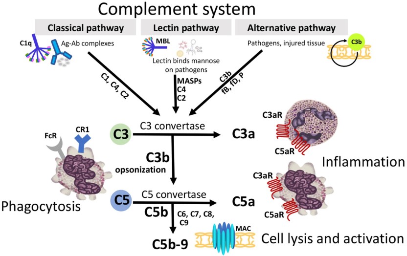 Figure 1. Common terminal pathway of three extracellular complement pathways
(Source: Girardi G, et al. 2020)
Complement activation is not restricted to the extracellular compartment, but can also occur intracellularly via cathepsin L, renin, thrombin, and fibrinolytic enzymes lysis of C3 and C5 and is secreted in an autocrine manner independent of the traditional convertase pathway. This activation pathway plays a key role in T cell homeostasis and effects. CTSL maintains a dynamic balance of quiescent CD4+ T cells by lysis of C3 to produce C3a and activation of C3aR on the lysosome, leading to low-level activation of mTOR. Migration of this intracellular C3 atypical activation pathway to the cell surface leads to activation of the TCR and the co-stimulatory molecule CD28. C3a and C3b trigger C3aR and CD46, respectively, to stimulate sustained activation of mTORC1. When CD4+ T cells are activated, CD46 triggers the intracellular lysis of C5 into C5a, which induces intracellular C5aR1 activation and contributes to C5aR1-dependent reactive oxygen species production.
The intracellular complement system is a relatively independent intracellular immune system that can regulate the function of the corresponding cells, and can be produced by a variety of cell types that can regulate cellular function within the cell. Unlike the extracellular complement system, which is traditionally synthesized and secreted by hepatocytes, ICS has been found in a wide range of cells other than hepatocytes, with intracellular complement components such as C3, C5, factor H (FH) and their corresponding receptors. The ICS is also named the composome because of its close association with the formation of nucleotide-binding oligomerization domain (NOD)-like receptor protein 3 (NLRP3) inflammatory vesicles.
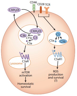 Figure 2. Noncanonical functions of intracellular complement
(Source: West EE, et al. 2018)
Intracellular C3 is mainly derived from receptor-mediated internalization of extracellular C3 hydrolysis products and self-synthesis of C3 proteins. Spontaneous hydrolysis of thioester bonds by C3 in the peripheral circulatory system occurs at a slow rate to form a C3 hydrolysis product, which enters the cytosol by binding to fibroblast lipoprotein receptor-related protein/α2-macroglobulin receptor (LRP/α2MR) and localizes to act in early endosomes. Cells can synthesize C3 proteins on their own, and some investigators have found that a signaling pathway mediated by the binding of human T-cell integrin lymphocyte function-associated antigen 1 (LFA-1) to endothelial cell-expressed intercellular adhesion molecule 1 (ICAM-1) induces T-cell C3 protein expression via activator protein 1 (AP-1).
Intracellular C3 plays an important role in maintaining CD4+ T cell homeostasis. Cellular homeostasis is maintained within resting T cells by maintaining basic cellular glycolytic metabolism, and when T cells are activated, C3b binds to CD46, which contains the functional end of the cytoplasmic terminus of CYT-1 on the surface of the T cell, and promotes differentiation of type I helper T (Th1) cells. When the T-cell antigen receptor (TCR) recognizes antigen and is activated, intracellular stores of C3aR and CD46 are rapidly translocated to the cell surface, while C3a and C3b are translocated extracellularly to bind to cell surface C3aR and CD46, respectively. Subsequent C3b-CD46 signaling drives GLUT-1 and L-type amino acid transporter 1 (LAT-1) expression and mediates upregulation of LAMTOR5 expression. Eventually large amounts of glucose and amino acids that are in-fluxed into the cell work together with LAMTOR5 to promote mTORC1 activation, causing γ-interferon (IFN-γ) secretion and Th1 cell formation.
Intracellular C3b is also involved in the cytotoxic effects of CD8+ T cells. When the organism is stimulated by relevant pathogens, CTSL rapidly lysis C3 to generate active C3b, which binds to CD46 on the cell surface via autocrine secretion and drives higher levels of fatty acid synthase, fatty acid-binding protein 5 expression, which in turn promotes the secretion of IFN-γ and granzyme B and induces cytotoxicity production. In addition, intracellular C3 is thought to be closely related to cancer development, and the increase in glycolysis resulting from its activation may be one of the mechanisms of tumorigenesis.
C5 is present in a variety of cells, but its origin is unclear. Similar to intracellular C3, activation of intracellular C5 in CD4+ T cells is also involved in Th1 cell formation. In conjunction with TCR and CD46, intracellular C5 is lysed into C5a and C5b, and C5a binds to C5aR1, which is only expressed intracellularly, inducing ROS generation. The production of endogenous ROS promotes the assembly of NLRP3 inflammatory vesicles and the secretion of IL-1β, which in turn maintains the Th1 response. When the activation signal is terminated, another C5a receptor, C5aR2, can form a dimer with C5aR1, thereby inhibiting the C5aR1-mediated extracellular signal-regulated kinase 1/2 (ERK1/2) signaling pathway and suppressing the Th1 effect, and the T-cells are then shifted from activated to resting state.
The function of the C5 system varies depending on the cell type. For example, in CD8+ T cells NLRP3 expression is not required for normal IFN-γ secretion and CTL activity, and in macrophages C5a-mediated signaling pathways even inhibit NLRP3 assembly and activation.
Since the complement system plays an important role in the body's defence against xenobiotics, removal of immune complexes from the body, and participation in acquired immunity, abnormalities in the complement system are closely related to autoimmune diseases, tumors, and a variety of other diseases. Decreased complement activity or dysfunction caused by defects in the complement system can lead to many serious diseases such as severe infections, systemic lupus erythematosus, kidney disease, and paroxysmal nocturnal haemoglobinuria.
Defects in some complement proteins, such as C2, C3, and MAC, increase the risk of infection in patients. The direct absence of C3 prevents the organism from acting as a regulator, thereby increasing the risk of infection with Gram-positive bacteria. The absence of MAC causes the body to be unable to attack the bacteria, resulting in gram-negative infections. The complement classical pathway is involved in the clearance of immune complexes and apoptotic cells and mediates complement-dependent B-cell tolerance, thus patients deficient in proteins such as C1, C2, and C3 are susceptible to autoimmune system diseases. Excessive complement activation caused by immune complexes in the renal vasculature can cause kidney damage and lead to glomerulonephritis. C3 and its regulatory proteins, factor I and factor H, are closely associated with the development of the disease. Deficiencies of factor I and factor H lead to disturbed C3 regulation. In animal models, C1q knockout mice have been found to produce autoantibodies that are not effective in clearing apoptotic cells and are therefore susceptible to immune complex-induced glomerulonephritis.
The complement system has long been considered important in the body's defence against tumors by promoting immune surveillance and inhibiting tumor progression. In concert with CD4+ T cells and antigen-presenting cells, complement promotes B-cell responses to soluble or membrane-expressed antigens from tumor cells and promotes the production of tumor antibodies. Complement can also generate MAC via the classical activation pathway leading to tumour cell lysis.
With further studies, researchers have found that the complement system and complement activation products can directly or indirectly promote tumor development. Imbalanced complement plays a key role in tumor promotion by maintaining local immunosuppression and chronic inflammation. C3aR and C5aR1-mediated signaling pathways have been found to promote the transformation of the tumor microenvironment towards favouring tumor progression by activating and polarising natural immune cells, suppressing effector T cells and releasing pro-tumor factors in animal models of cervical, colorectal and lung cancers. On the other hand, C3 plays an important role in the pre-metastatic tumor microenvironment and can promote tumor cell colonisation in metastatic target organs. Complement also induces metalloproteinases, increases the expression of stress fibres and filamentous fibres and degrades the extracellular matrix thereby regulating tumor cell motility and invasion. In addition, complement is associated with angiogenesis and tumorigenesis. C3a and C5a upregulate the expression of vascular endothelial growth factor (VEGF) and basic fibroblast growth factor (BFGF), thereby promoting endothelial cell proliferation.
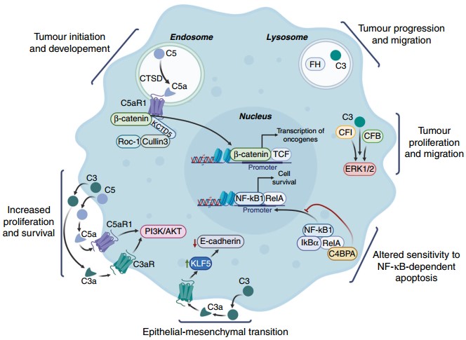 Figure 3. Schematic representation of roles for intracellular complement proteins in cancer cells
(Source: O'Brien RM, et al. 2023)
References
| Target | Cat. No. | Product Name | Size | Species Reactivity | Application | Detection Sample | |
| C1q | DEIA3234 | Human C1q-Ab(Anti-Complement 1q Antibody) ELISA Kit | 96T | Human | Quantitative | Serum, plasma, tissue homogenates and other biological fluids | Inquiry |
| DEIASL233 | Human Complement 1q ELISA Kit | 96T | Quantitative | Serum, plasma, tissue homogenates and other biological fluids | Inquiry | ||
| C3 | DEIA7754 | Guinea pig C3(Complement Component 3) ELISA Kit | 96T | Guinea pig | Quantitative | Serum, plasma, tissue homogenates and other biological fluids | Inquiry |
| DEIA8289 | Mouse CFB(Complement Factor B) ELISA Kit | 96T | Mouse | Quantitative | Serum, plasma, tissue homogenates and other biological fluids | Inquiry | |
| c4 | DEIA6349 | Human Complement C4a des Arg ELISA Kit | 96T | Human | Quantitative | EDTA plasma | Inquiry |
| DEIA6798 | Human C4A(Complement C4-A) ELISA Kit | 96T | Human | Quantitative | Serum, plasma, tissue homogenates and other biological fluids | Inquiry | |
| C5 | DEIA-BJ2734 | Guinea pig Terminal complement complex C5b-9 ELISA Kit | 96T | Quantitative | Serum, plasma, cell culture supernatants, body fluid and tissue homogenate | Inquiry | |
| DEIA-BJ2847 | Porcine Terminal complement complex C5b-9 ELISA Kit | 96T | Quantitative | Serum, plasma, cell culture supernatants, body fluid and tissue homogenate | Inquiry | ||
| DEIA-BJ2881 | Canine Terminal complement complex C5b-9 ELISA Kit | 96T | Canine | Quantitative | Serum, plasma, cell culture supernatants, body fluid and tissue homogenate. | Inquiry | |
| C5B-9 | DEIA-BJ2053 | Rat Plasma levels of terminal complement complex ELISA Kit | 96T | Quantitative | Serum, plasma, cell culture supernatants, body fluid and tissue homogenate | Inquiry | |
| DEIA-BJ2758 | Porcine Plasma levels of terminal complement complex ELISA Kit | 96T | Quantitative | Serum, plasma, cell culture supernatants, body fluid and tissue homogenate | Inquiry | ||
| CR1 | DEIA-BJ433 | Human Complement fragment 3b rccptor ELISA kit | 96T | Quantitative | Serum, plasma, cell culture supernatants, body fluid and tissue homogenate | Inquiry | |
| CFB | DEIA-BJ2389 | Mouse Complement Factor B ELISA Kit | 96T | Quantitative | Serum, plasma, cell culture supernatants, body fluid and tissue homogenate | Inquiry | |
| DEIA-BJ2765 | Porcine Complement Factor B ELISA Kit | 96T | Quantitative | Serum, plasma, cell culture supernatants, body fluid and tissue homogenate | Inquiry |
| Target | Cat. No. | Product Name | Expression System | Tag/Conjugate | Application | |
| C1q | DAG-WT2554 | Complement C1q control | N/A | Unconjugated | Immunoassays | Inquiry |
| C3 | DAG-WT2115 | Recombinant Human Complement component 3 Protein [His] | HEK293 cells | His | Immunoassays | Inquiry |
| DAG-WT2552 | Complement C3 control | N/A | Unconjugated | Immunoassays | Inquiry | |
| C4 | DAG-WT2553 | Complement C4 control | N/A | Unconjugated | Immunoassays | Inquiry |
| CFD | DAG-WT2069 | Recombinant Human Complement Factor D [hFc] | HEK293 cells | hFc | Immunoassays | Inquiry |
| CFP | DAG4680 | Human Complement Factor P | N/A | Unconjugated | N/A | Inquiry |
| Target | Cat. No. | Product Name | Host | Isotype | Application | |
| C3 | CABT-B023 | Anti-Complement C3 monoclonal antibody, clone 20D8 | Mouse | IgG1 | FC, ELISA | Inquiry |
| CABT-B024 | Anti-Complement C3 monoclonal antibody, clone 2I9 | Mouse | IgG1 | IF, ELISA | Inquiry | |
| CABT-B025 | Anti-Complement C3 monoclonal antibody, clone 7D0 | Mouse | IgG1 | FC, ELISA | Inquiry | |
| CABT-B026 | Anti-Complement C3 monoclonal antibody, clone 9F22 | Mouse | IgG1 | ELISA | Inquiry | |
| CABT-B027 | Anti-Complement C3b-iC3b monoclonal antibody, clone 4F8 | Mouse | IgG1 | FC, ELISA | Inquiry | |
| CABT-B028 | Anti-Complement C3b-iC3b monoclonal antibody, clone 6H0 | Mouse | IgG2a | Neut, FC | Inquiry | |
| CABT-B029 | Anti-Complement C3b-iC3b monoclonal antibody, clone 8D23 | Mouse | IgG1 | ELISA | Inquiry | |
| c4 | DPAB1836 | Magic? Anti-C4c Complement polyclonal antibody | Rabbit | IgG | IHC | Inquiry |
| C5 | DPATB-H81286 | Anti-E. coli Complement 5 Polyclonal antibody | Rabbit | IgG | IHC-FoFr, WB, IHC-Fr | Inquiry |
| C5AR1 | CABT-L3174 | Mouse Anti-Human Complement Component C5a R1 monoclonal antibody, clone 458325 | Mouse | IgG2a | FC, Neut | Inquiry |
Loading ......
