Introduction of Equine ELISPOT Assay
The vast majority of ELISPOT assays is performed on human and mouse or rat immune system cells, with far fewer assays of primate, feline, and canine cells. Because of the valuable and unique information that can be collected using ELISPOT assays, however, they can also be utilized to study many other species. Cells from both domesticated livestock and wild animals can be tested to monitor their health and to diagnose immune system status. Until fairly recently, most studies evaluating cytokine induction in horses have relied on bioassays, ELISA, or methods assessing mRNA expression. The development of a cell-based immunoassay, such as ELISPOT, has been delayed due to the lack of species-specific antibodies and cytokine standards. To bridge the gap and to expand the utility of ELISPOT assays for equine research, we have developed antibodies against equine cytokines and designed ready-to-use ELISPOT kits to study the frequency of IFNG and IL-4 secretion from equine peripheral blood mononuclear cells (PBMCs) in a multidonor study. This chapter describes an equine ELISPOT protocol, and discuses important details for the performance of a successful assay.
Methods of Equine ELISPOT Assay
Isolation of Equine Peripheral Blood Lymphocytes
- Collect blood samples from healthy donors in sodium heparin. Centrifuge whole blood in a 50-mL centrifuge tube at 500 × g for 10 min.
- Discard upper plasma layer after centrifugation, and refill 50-mL tube with sterile PBS. Using density centrifugation separation, layer 25 mL of blood/PBS mixture on 20 mL of 1.077 g/mL Ficoll-Paque™ PLUS at 25°C (see Note 3) and centrifuge at 500 × g for 30 min.
- Discard the top layer after centrifugation and transfer PBMCs (buffy coat layer) into two sterile 50-mL tubes.
- PBMCs are then resuspended in 45 mL of sterile PBS and centrifuged for 5 min at 500 × g.
- Discard supernatant and resuspend the pellet in 10 mL of red blood cell lysing solution and incubate for 5 min at room temperature.
- After lysing, add sterile PBS to reach 50-mL graduation mark on the tube to resuspend PBMCs.
- Centrifuge tubes for 5 min at 500 × g.
- Discard supernatants and add 30–40 mL of RPMI complete to the tubes with PBMCs.
- Mix a small sample of cells 1:2 with trypan blue dye and pipette 10 ML of this mixture into each side of a hemacytometer under a coverslip (see Note 4). Count cells under the microscope using 20× lens and phase-contrast condenser.
ELISPOT Assay
- Plate PBMCs (100 ML/well; six wells per group) into the ELISPOT plates at cell concentrations of 105 and 106 cells/ mL (see Notes 5 and 6).
- Stimulate PBMCs with either 0.5 Mg/mL of calcium ionomycin and 50 ng/mL of PMA or 4 Mg/mL of Con A added directly to cells in ELISPOT plates and incubated in a CO2 incubator at 37°C for 18 h (see Notes 7 and 8).
- After finishing the incubation, aspirate PBMCs from the plates and wash the plate by rinsing wells four times with wash buffer (see Notes 9 and 10).
- Make working solutions of detection antibodies by diluting concentrated detection antibody 1:120 with dilution buffer.
- Add 100 ML of detection antibody working solution into each well and incubate ELISPOT plates overnight at 4°C.
- Wash plates three times with the wash buffer.
- Prepare working solution of streptavidin–alkaline phosphatase by mixing the concentrated stock solution 1:120 with corresponding dilution buffer.
- Add 100 ML of streptavidin–alkaline phosphatase working solution into each well and incubate for 2 h at room temperature.
- Wash plates three times with wash buffer.
- Add 100 ML of ready-to-use BCIP/NBT substrate into each well and incubate for 30–60 min at room temperature in a place protected from direct light.
- Wash plates three times with distilled water and let them dry completely (see Note 11).
- Quantify spots using automated ELISPOT reader.
Our study demonstrated that Con A had a more profound stimulating effect than CaI + PMA on IL-4 secretion from equine PBMCs, whereas both stimulating and inhibitory effects were observed on PBMCs from the same animals for IFNγ secretion.
Con A in general had a stronger stimulating effect on secretion of IFNγ and IL-4 than CaI + PMA, but the responses can vary significantly among individual donors.
Our study demonstrated that ELISPOT assay can be successfully used to determine the frequency of cytokine-secreting equine PBMCs. We have also shown that ELISPOT is a very convenient and easy-to-use assay capable of covering a large dynamic range in IFNγ and IL-4 secretion among different donors which may reflect animal health conditions and/or serve as a valuable diagnostic tool.
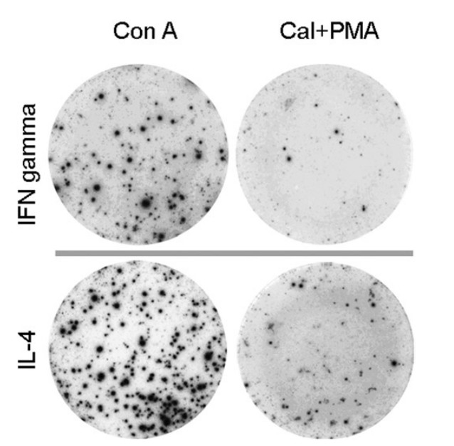 Figure 1. Typical ELISPOT images of equine IFNG and IL-4.

 Loading ......
