
Inquiry Basket
 Loading ......
There are two different types of membranes (in 96-well plate formats) that are used in ELISPOT applications: Nitrocellulose (NC) and polyvinylidene fluoride (PVDF) with PVDF becoming increasingly dominant over the past 10 years (although some features and guidance on how the use of NC plates will be provided, the emphasis in this chapter will be on PVDF and the use of PVDF-based devices in ELISPOT). The reason that NC and PVDF are the principal membrane types used in ELISPOT is based largely on their fortuitous suitability in a range of non-ELISPOT applications rather than on the development of optimized assay substrates to address the specific needs of the ELISPOT assay. The chronology of relevant membrane and application developments is outlined.
1954: NC membranes become commercially available. Principal use is in removing bacteria from aqueous samples (sterile filtration).
1975: 0.45μm NC membrane used in DNA hybridization (Southern Blotting) assay.
1975: PVDF membranes become commercially available. Initially, principal use is in removing bacteria from aqueous samples. PVDF is more durable and more solvent resistant than NC and as such is more suited to large-scale filtration applications.
1979: 0.45μm NC membrane used in immunodetection of electro-blotted proteins.
1983: ELISPOT assay developed on plastic, 96-well plates.
1985: 96-well plate with NC membrane becomes commercially available. Intended application is dot blotting of nucleic acids using vacuum transfer (instead of capillary transfer).
1986: 0.45μm PVDF membrane used in Western Blotting Assay.
1988: 96-well plate with NC membrane used in ELISPOT.
1992: 96-well plate with PVDF membrane becomes commercially available. Intended application is dot blotting of proteins using vacuum transfer (instead of electro-blotting). Millipore Corporation reports in its 1994/5 Catalog that this plate can be used in ELISPOT.
1995: Articles citing improved ELISPOT results using PVDF plates are published.
2002: Automation compatible 96-well plates become commercially available making it possible to use robotic liquid handlers to perform many of the reagent additions and plate washing steps associated with high throughput screening (HTS).
2003: Automation compatible, 96-well plates designed specifically for ELISPOT becomes commercially available.
2003: Membrane-bottomed, 8-well strip plates become commercially available for diagnostic and research ELISPOT applications.
2005: PVDF membranes are made with low background fluorescence making it much more possible to perform fluorescent ELISPOT assays.
2008: Patented T-Spot ELISPOT assay receives FDA PMA approval as a clinical diagnostic test for TuberculosisAs.
NC and then PVDF membranes were first developed to serve the needs of sterile filtration applications. In ways that were never anticipated by membrane manufacturers, the porosity and binding properties of these membranes enabled them to be used in two extremely important and burgeoning research applications; nucleic acid hybridization assays and Western blotting. Eventually, to serve the specific requirements of molecular biology and protein chemistry applications, NC and PVDF membrane-bottomed 96-well plates were developed and made commercially available. Independently and separately, ELISPOT assays were developed on 96-well plastic plates and took advantage of enzyme-linked immunosorbent assay (ELISA) techniques that had been perfected in that format. Since the immunodetection component of ELISPOT assays and Western blotting is essentially identical, it was only a matter of time until the overwhelming majority of ELISPOT assays were performed on membrane-bottomed, 96-well plates.
PVDF membranes were developed in part to overcome major limitations of NC, including poor chemical compatibility, shedding of particulates, and brittleness. Many of the same companies that were able to produce NC also developed the ability to make PVDF membranes over a range of different pore sizes. The PVDF polymer itself is highly resistant to chemical degradation – except in the presence of strong alkali (pH greater than 12) – and membranes made from PVDF are sufficiently elastic to withstand a broad range of fabrication conditions (including sonic welding and pleating) and high-pressure filtration applications. Millipore, an early provider of PVDF membranes, created the trademark, "Durapore®" to call attention to these attributes. PVDF, like NC, is intrinsically hydrophobic. Whereas NC is only marginally hydrophobic and can be made water wettable by adding a surfactant (e.g., glycerin) or detergent (e.g., Triton®-X 100) to the membrane, PVDF is extremely hydrophobic and requires significant surface modification to make it compatible with aqueous solutions (see Note 1). A considerable amount of chemistry and patented technology was subsequently developed to make PVDF membranes hydrophilic. The important point to note is that even though the polymer itself is very hydrophobic, membranes made from PVDF can be either hydrophilic or hydrophobic depending on whether the manufac turer has modified or covered over the polymer surface with a secondary chemical treatment that is water compatible.
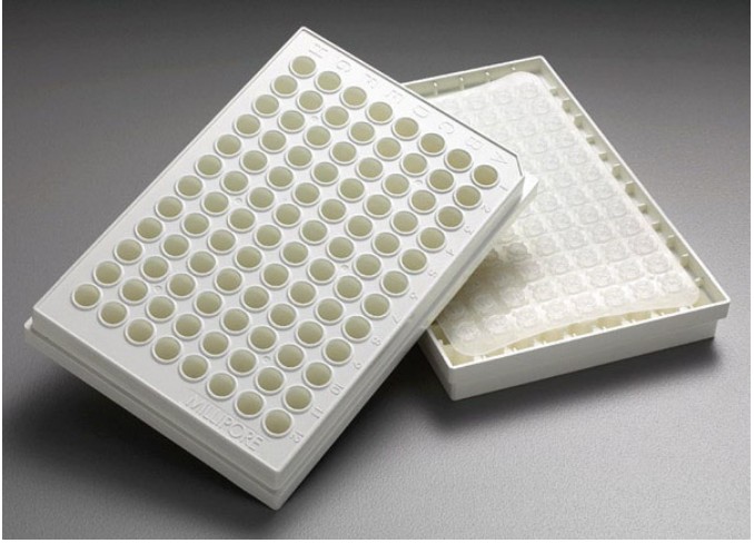 Figure 1. 96-well filter-bottomed plate used for ELISPOT and HTS applications.
The discussion of PVDF is relevant to ELISPOT in that the hydrophobic version of the 0.45 μm membrane was found to be an excellent Western blotting substrate. PVDF binds proteins by hydrophobic interactions (van der Waal's forces). This applies, of course, only to hydrophobic PVDF membranes. Most types of hydrophilic PVDF will not bind proteins to any appreciable degree. Interestingly, hydrophobic PVDF will bind single-stranded DNA and RNA, but will not bind double-stranded DNA.
The use of PVDF membranes in Western blotting type applications grew rapidly. Unlike NC membranes, PVDF membranes could stand up to automated protein sequencing chemistries and were better able to retain low molecular weight proteins and peptides. PVDF was also better suited to a wider range of detection techniques, including fluorescence and chemiluminescence. One significant drawback of PVDF is the need to pre-wet the membrane in an alcohol solution prior to using it in Western blotting and most other applications that include immunodetection. The requirement to pre-wet with alcohol – which is completely necessary in Western blotting applications – is not universally applicable in ELISPOT. Differences in ELISPOT protocols with regard to the pre-wetting step are significant and are likely to have an impact on the performance of the assay. This topic is discussed in greater detail later in this chapter (see Note 2).
The rationale behind the development and commercialization of NC and PVDF is clear from the perspective of filtration applications. As has been pointed out already, the use of each of these membrane types in molecular biology and protein chemistry applications was based on a fortuitous combination of membrane properties; high permeability (due to high porosity), and high DNA/ RNA and protein binding. The development of 96-well PVDF and NC bottomed plates was primarily driven by the secondary (i.e., biochemistry) applications. One of the attributes of both Southern and Western blotting is that (DNA) hybridization and antibody binding are diffusion limited reactions. In other words, the reactant in solution (complimentary DNA or antibody) must diffuse to the surface of the membrane before it can couple with the immobilized reactant (DNA or protein). In these types of solid phase reactions, the times required to reach equilibrium binding are much longer as compared to reactions in which both reactants are in solution. Typically, DNA hybridization (Southern blotting) and immunodetection (Western blotting) require from 2 to 24 h. Membrane-bottomed, 96-well plates, made it possible to reduce the time requirements associated with solid phase binding. (Earlier, simpler versions of these plates, called "Dot Blot" or "Slot Blot" apparatuses provided the first opportunity to exploit the benefits of filtration in these applications). Filtration of the reactant in solution through the membrane brings it into intimate contact with the reactant immobilized on the membrane surface. So long as the filtration rate is kept low enough for hybridization or binding to take place, the time required to achieve efficient capture can be dramatically reduced. Additionally, all wash steps in between various reactions can be accomplished using filtration. The development of membrane-bottomed plates in conjunction with compatible vacuum manifolds made it possible to carry out from 1 to 96 different hybridization or immunodetection assays using less reagents and requiring less time as compared to standard methodologies.
In addition to the membranes and other features that were useful for these applications, the plates were also designed to allow for discrete liquid transfer from the top (membrane-containing) plate to a (standard, plastic, 96-well) receiver plate. The filter plate components that allow for this to occur have the potential to interfere with ELISPOT applications in at least three different ways.
Preventing these types of problems during the ELISPOT assay and optimizing the different parts of the protocol are reviewed elsewhere.
There are other plate attributes that may have an impact on ELISPOT assay performance or analysis. The membrane inside each well needs to be planar within a fraction of a millimeter (e.g., ±0.1 mm) in order to provide the best assay results. Lack of membrane flatness may contribute to difficulties in imaging depending on the type of microscopy being used. Additionally, if the membrane is bowed, cells may tend to settle unevenly or roll to the relative low points (often either the center or the periphery) during incubation. Consequently, spots may become very unevenly distributed and difficult to enumerate accurately especially if there is any spot confluence. The plate itself should also be flat (within a tolerance of perhaps 1 mm corner to corner) to assure compatibility with plate washers and most of the imaging software that supports automated image acquisition and analysis. There is one other feature of 96-well plates that has the potential to introduce variability into the ELISPOT assay. The 96-well plate is arrayed as 8 rows of 12 wells. Wells at the periphery of the plate are fundamentally different from "interior" wells insofar as they are in the most direct contact with the plate surroundings. Depending on incubation conditions and other protocol steps, this physical distinction may have some impact on one or more parts of the ELISPOT assay (see Note 3).
ELISPOT assays were first developed on plastic, 96-well plates. Shortly after NC-bottomed filter plates became available, the majority of ELISPOT assays were carried out in those plates. When PVDF filter plates were introduced, some investigators chose to use PVDF plates and some continued to use NC. The reasons for choosing one plate (membrane) over the other are highly varied and will not be addressed in detail here although over the past 10 years, the percentage of ELISPOT assays performed on PVDF plates has steadily increased. The fact that some laboratories and individual researchers feel strongly that one membrane is superior to the other runs contrary to the large body of Western blotting experience: Despite some clear-cut differences in how each of the membranes can be used, there is essentially no reported difference in terms of detection sensitivity or signal to noise on NC versus PVDF in the Western blotting application. This having been said, it is clear that the two membranes and their properties are quite different.
Like NC, and for the same reasons, the PVDF membrane in plates used for ELISPOT has a nominal pore size of 0.45 Mm. PVDF is nominally 135 Mm thick and about 65–70% porous. The BET surface area is about the same as NCs and its surface area ratio is somewhat higher – around 350 (see Note 4).
Consequently, PVDF can bind upward of 350 Mg/cm2 of IgG or in excess of 100 Mg per well of a 96-well plate. As with NC, blocking of PVDF should occur within a few hours (or less) of antibody coating. Failure to do so may result in a rapid and significant loss of antibody activity. Once antibody has been coated and the membranes have been blocked (and washed using deionized water or very low molarity buffer), plates can be stored (desiccated and at room temperature) for weeks or even months (see Note 5). PVDF that has been coated with protein (e.g., as a consequence of antibody coating and blocking) will rewet spontaneously upon the addition of aqueous media.
The major difference between NC and PVDF in ELISPOT applications is related to their mechanisms of binding and associated differences in handling or pretreatment. PVDF is very hydrophobic (its surface energy is approximately 21 dyn/cm) and will not wet out in water. In Western blotting applications, PVDF is always pre-wet in alcohol (typically 50–100% methanol), then normally exchanged in water, and ultimately equilibrated in a (transfer) buffer solution before applying the membrane to the polyacrylamide gel for electrophoresis. The fact that hydrophobic PVDF membranes will not wet out spontaneously in water – unless coated by (blotted) proteins – is even exploited in a Western blotting application called "Transillumination". The overwhelming majority of literature in Western blotting references the pre-wetting step, so it is not a surprise that many ELISPOT protocols also include an alcohol pre-wet step. What is surprising is that some ELISPOT protocols do not include a pre-wet step. At Millipore, experiments were performed to determine the relative performance PVDF 96-well plates that were either pre-wet with 15 MmL of 70% v/v methanol in water and rinsed, or not pretreated at all prior to antibody coating. Briefly, following the alcohol pretreatment (or no pretreatment), plates were coated with 1 Mg of anti-human interferon-gamma, and blocked for 2 h in tissue culture media containing 10% fetal bovine serum. 50,000 peripheral blood mononuclear cells (see Note 6) were added per well to 16 wells per plate, stimulated with 0.5 μg phytohemagglutinin and the plates were incubated overnight in a humidified, 37°C, 5% CO2 tissue culture incubator. ELISPOTs were visualized using biotinylated anti-human interferon-gamma, conjugated avidin-alkaline phosphatase and BCIP/NBT Plusand then enumerated using an automated microscope and its associated software.
In these experiments, the cells in the untreated (nonpre-wet) plates produced approximately 30% fewer detectible spots. However, the consistency well to well and plate to plate was equivalent. Spot quality (intensity, uniformity, and size) and overall assay background were comparable in both plate types. Considering that half these results were obtained without pre-wetting, the comparable, side-by-side performance is quite remarkable. It would appear that the determination to pre-wet with alcohol or not can be made by individual laboratories based on their reagent selections and particular assay requirements.
PVDF, like NC, is fully compatible with ELISA detection involving precipitating, color-forming substrates and chemiluminescent substrates. Although the fluorescence background of PVDF is also high, due principally again to light scattering, ELISPOT assays have been developed on PVDF that are based on the use of fluorescently labeled antibodies. The ability to develop fluorescent immunoassays on PVDF membranes – particularly useful for applications in which more than one antigen is being simultaneously detected – was significantly enhanced with the commercialization of a specialized PVDF membrane that was specifically developed for fluorescence detection.
As was stated earlier in the chapter, 96-well plates were initially designed to enable sample filtration and discrete transfer of fluids. These design elements, which are not useful for ELISPOT, can actually interfere with the ELISPOT assay if the user is not careful. Consequently, in 2003, a 96-well plate with PVDF was commercialized that was designed specifically for ELISPOT applications. There is no underdrain to facilitate discrete fluid transfer in these ELISPOT-specific plates. Consequently, problems associated with alcohol pre-wetting and membrane removal after ELISPOT development are virtually eliminated. One of the issues with 96-well plates of all types is that many ELISPOT assays do not end up requiring the use of all 96-wells and unused wells essentially get wasted. This is especially true in diagnostic applications in which only one or two patients may be tested at a time. For these types of low-throughput applications, there are now 8-well strip plates available. Individual (8-well) strips can be antibody coated, blocked, and used in an assay without any impact on the remaining strips. This particular format was first used in a diagnostic test approved by the FDA in 2008 for tuberculosis infection and is now also commercially available for research and development applications.
Although with proper optimization, it is likely that comparable ELISPOT performance can be achieved using either type of membrane, it is unlikely that the same protocol will work equally well in both cases. Neither PVDF nor NC is a drop-in for the other (see Note 7). Fundamental differences in the two membranes, especially with regard to their mechanisms of binding and their ability to wet directly in water will affect their behavior in ELISPOT. Modifications that have been made in the design of 96-well plates to make them compatible with automation – including stricter dimensional specifications and rigid sidewalls (to allow handling by laboratory robotics and provide space for bar codes) have also benefited ELISPOT applications. These plates are now fully compatible with standard plate washers as well as with imaging equipment and image analysis software. It is reasonable to believe, based on the importance of ELISPOT and the impressive growth in the number of assays being performed, that membranes and membrane-based plates may someday be optimized specifically for this application.
Loading ......
