
Inquiry Basket
 Loading ......
The enzyme-linked immunosorbent spot (ELISpot) assay is an ELISA-based method commonly used for the identification and enumeration of cytokine-producing cells with exquisite sensitivity at the single-cell level.
One of the most common applications of the assay is the enumeration of IFNγ-secreting cells in response to antigenic stimulation. Typically, the assay is performed by stimulating antigen-specific T cells within peripheral blood mononuclear cells (PBMCs) in wells coated with an antibody that binds to the desired cytokine. In response to the recognition of the antigen, T cells specifically release IFNγ, which is then trapped to the anti-IFNγ antibody. After a washing step, which removes the cells from the wells, the location of the cytokine-releasing cell is visualized by an enzyme-labeled detection antibody and its corresponding chromogenic substrate, which has to be non-soluble to become attached to the surface of a well. The end result is a set of colored spots, each of which represents an area where a cell secreting IFNγ had been located.
T cells present among the PBMC recognize peptides presented on the HLA surface of antigen-presenting cells (APC) like monocytes and macrophages also present in the PBMC sample. Whereas HLA-binding oligopeptides can be added directly to the ELISpot assay without the necessity of a preincubation step, complex protein antigens must be internalized and processed by professional antigen-presenting cells (APC) for efficient presentation via HLA class I or II. To overcome this issue, we have developed a serum-free DC-ELISpot protocol using standardized DC for sensitive enumeration of T cells recognizing protein antigens, based on the assay described by Nehete and colleagues. In this variation of the ELISpot assay, the stimulation is performed with autologous DC previously loaded with the desired protein antigen.
Here we describe the standard and the DC-ELISpot assay protocol for the detection of IFNγ-secreting cells upon antigen stimulation. For measuring immune responses against oligopeptide antigens, a direct ELISpot protocol may be used; however, when the target antigen is a protein, the DC-ELISpot should be the preferred protocol.
Although the assay is described to detect IFNγ-secreting cells, it can be easily adapted for the detection of other cytokines providing that the appropriate capture and detection antibody pair is available.
Prepare all solutions under sterile conditions; use cell culture-grade and analytical-grade reagents as required. Prepare and store all reagents at 4 °C (unless otherwise indicated). Diligently follow all waste disposal regulations when disposing waste materials. Avoid the use of sodium azide.
Cell Culture Reagents
1. Serum-free ELISpot cell culture medium: CellGro DC Medium supplemented with penicillin/streptomycin. Keep sterile and store at 4 °C.
2. PMA/ionomycin solution: requires phorbol 12-myristate 13-acetate (PMA, cell culture grade, Sigma-Aldrich) and ionomycin (cell culture grade, Sigma-Aldrich). Prepare a fresh solution containing 10 ng/ml PMA and 4 μg/ml ionomycin in the ELISpot cell culture medium.
ELISpot Assay Reagents
3. Dulbecco's phosphate-buffered saline (DPBS) without calcium and magnesium. Keep sterile and store at room temperature.
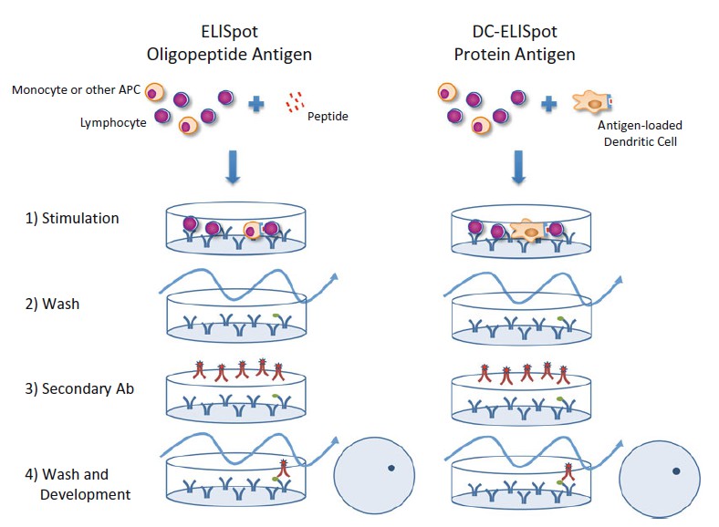 Figure 1. Schematic overview of the ELISpot assay (left ) and the DC-ELISpot variation ( right ).
4. Serum-free ELISpot medium: CellGro DC Medium supplemented with penicillin/ streptomycin. Keep sterile and store at 4 °C.
5. 96-well MultiScreen plate, hydrophobic with PVDF membrane.
6. Human IFNγ ELISpot pair.
7. ELISpot washing buffer: DPBS containing 0.005 % Tween 20.
8. Streptavidin-alkaline phosphatase enzyme conjugate, 1:1,000 dilution in DPBS.
9. AP Conjugate Substrate Kit.
10. Dilute 25× AP color development buffer 1:25 in water (400 μl AP + 9,600 μl H2O). Buffer can be distributed in aliquots and stored at 4 °C.
Equipment
11. Laminar flow cabinet.
12. CO2 cell culture incubator.
13. Automated ELISpot reader.
Equipment
This assay makes use of life cells; therefore, the processing of samples and/or performing cell culture-based assays on clinical specimens from patients and animals must be performed in the appropriate biosafety level (BSL) laboratory (BSL-2 or above depending on the type of sample). Laboratory safety procedures include at least all standard BSL-2 procedures with enhancements as follows: (1) laboratory personnel have specific training in handling pathogenic and potentially lethal agents and are supervised by competent scientists who are experienced in working with these agents; and (2) the laboratory should have special engineering and design features to ensure directional airflow from clean to potentially contaminated areas. Supervisors are responsible for ensuring that technicians are properly trained to work safely in the laboratory.
All these steps must be performed under sterile conditions in a laminar flow cabinet. Keep the cells on ice during preparation to prevent unspecific activation.
All these steps must be performed under sterile conditions in a laminar flow cabinet. Block Membrane with Serum-Free ELISpot Medium
1. Decant primary antibody solution.
2. Wash off unbound antibody with 150 μl sterile DPBS, decant wash, and repeat.
3. Block membrane with 150 μl/well of serum-free ELISpot medium for at least 2 h at 37 °C.
Cell Culture and Stimulation
4. Stimulation: Plate 100 μl/well of the rested PBMC suspension (1–4 × 10⁶ viable cells/ml = 1–4 × 10⁵ viable cells/well) together with 100 μl/well of the peptide diluted in serum-free ELISpot medium at a concentration of 1–10 μg/ml in at least three replicates. If the stimulator is a protein, add instead 100 μl/ well of antigen-loaded DC suspension (1–10 × 104 viable cells/ ml = 1–10 × 103 viable cells/well) (see Notes 4 and 5).
5. Background control: Plate 100 μl/well of the rested PBMC suspension (1–4 × 10⁶ viable cells/ml = 1–4 × 10⁵ viable cells/ well) together with 100 μl/well of serum-free ELISpot medium. If the stimulator is a protein, add instead 100 μl/well of non- loaded DC suspension (1–10 × 104 viable cells/ ml = 1–10 × 103 viable cells/well).
6. As positive control, plate 10 × 103 PBMC/well triplicate in 150 μl ELISpot-incubation medium + 50 μl PMA/ionomycin solution.
7. Incubate overnight at 37 °C 5 % CO2.
Important: Do not disturb the plate during the incubation period or artifacts may form.
The following steps do not require sterile conditions. Use appropriate BSL-2 biosafety procedures
Secondary Antibody
1. Flick and blot to decant cells (see Note 6).
2. Wash the plate six times with ELISpot washing buffer.
Important: The ELISpot washing buffer is DPBS with 0.005 % Tween 20. It is important not to exceed 0.01 % Tween 20 to prevent the possibility of leakage and artifacts.
3. Dilute 2 μg/ml biotinylated anti-IFNγ antibody in DPBS/0.5 % human serum albumin and add 100 μl/well.
4. Incubate for 30 min at 37 °C, 5 % CO2, and 95 % humidity (or alternatively at 4 °C overnight).
5. Wash the plate six times with ELISpot washing buffer.
Enzyme Conjugate and Substrate Development
6. Prepare streptavidin-alkaline phosphatase enzyme conjugate at 1:1,000 dilution in DPBS.
7. Add 100 μl per well of the diluted streptavidin-alkaline phosphatase. Incubate for 30 min at room temperature in the dark (cover the plate with aluminum foil) (see Note 7).
8. Decant streptavidin, and wash three times with ELISpot washing buffer, followed by four washes with DPBS without Tween 20 (see Note 8 ).
9. Dilute 25× AP color development buffer 1:25 in water (400 μl AP + 9,600 μl H 2 O).
10. Add 100 μl of reagent A and 100 μl of reagent B to 10 ml of the previous solution.
11. Add 100 μl/well of development solution.
12. Incubate in the dark until it is possible to see the spots in the positive control wells for 7–10 min (see Note 9).
13. Stop development under running water and wash extensively. While washing, remove the underdrain and continue rinsing.
14. Let plate dry in the dark.
Plate Reading
15. Read the plates in an automated ELISpot reader; the reading process is performed in a semiautomated process. Trained personnel should make adequate adjustments for technical artifacts and audit the read process.
Reference
Loading ......
