Introduction of Correlative Microscopy Protocol
The main challenge in immuno-EM is the retention of antigenicity while preserving fine ultrastructure. The prerequisite to successful application of the method is the quality of the antibody in terms of good affinity and specificity for native proteins. Applicability of an antibody for western blot is not sufficient since this method detects denatured proteins. With immuno-EM the antibody has to recognize its epitope after aldehyde fixation and resin embedding which can remove up to 90 % of the epitopes. The first step in the method is to try the immunolabeling at the light microscopy level to test the antibody labeling under fixation conditions suitable for EM. Once the labeling obtained is satisfactory, the protocol can be adjusted to reach a compromise between preservation of ultrastructure and labeling. One of the advantages of pre-embedding immuno-EM is that the labeling is done before the embedding, thereby avoiding the further loss of antigenicity during this step.
The development of FluoroNanogold (FNG) provided a marker system with a greater permeability and labeling sensitivity. These probes comprise a 1.4 nm gold particle (Nanogold) conjugated to a Fab’ fragment and a fluorescent label, allowing for imaging by correlative light and electron microscopy. Particularly, the combination of FNG with an intensification step (silver or gold intensification) enhanced the detection of intracellular antigens at the EM level by using pre-embedding methods.
The ability to track the distribution of macromolecular complexes by utilizing multiple imaging modalities is a valuable tool in biology and sparked a general interest over the years to develop various approaches for correlated/correlative microscopy. Current techniques include plastic embedding compatible with fluorescence preservation. Particularly, some hydrophilic resins are capable of low temperature polymerization, thereby avoiding denaturing proteins due to heat or stringent dehydration.
In this chapter we apply this method to visualize connexin43 proteins endogenously expressed in normal rat kidney (NRK) cells. With the use of LR White resin we were able to image the fluorescent signal from Cx43 proteins labeled with FNG in plastic, followed by the visualization of their subcellular localization by EM.
Methods of Correlative Microscopy Protocol
Perform a fixation series of paraformaldehyde and glutaraldehyde dilutions, and assess where the fluorescence signal is lost.
- Remove the culture medium from the MatTek dishes containing the cells and wash three times with HBSS pre-warmed up at 37 °C. Fix cells with pre-warmed fixative at 37 °C (4 % PFA, 4 % PFA + 0.1 % glut, 2 % PFA, 2 % PFA + 0.1 % glut) for 5 min at room temperature followed by 30 min over ice. All solutions and steps from this point on are utilized and performed at 4 °C for the preservation of the ultrastructure (see Note 1).
- Remove fixative and wash cells three times for 2 min each in ice-cold 1× PBS. Note: during any washing step the cells should never become dry to avoid irreversible damage to morphology.
- Incubate in blocking buffer for 1 h at 4 °C (see Note 2).
- Incubate in primary antibody diluted in blocking buffer at 4 °C for 1 h on a rocking platform set for gentle agitation (see Note 3).
- Rinse cells with Working Buffer, six times for 2 min each at 4 °C.
- Incubate in secondary antibody diluted in blocking buffer at 4 °C for 1 h on a gentle rocker, protected from light. Remember: keep cells in the dark from now on, to preserve fluorescence.
- Rinse cells with Working Buffer, six times for 3 min each at 4 °C.
- Wash cells with ice-cold 1× PBS, three times for 3 min each at 4 °C.
- Check staining with a fluorescence microscope; proceed with EM processing only if good signal and specificity is observed.
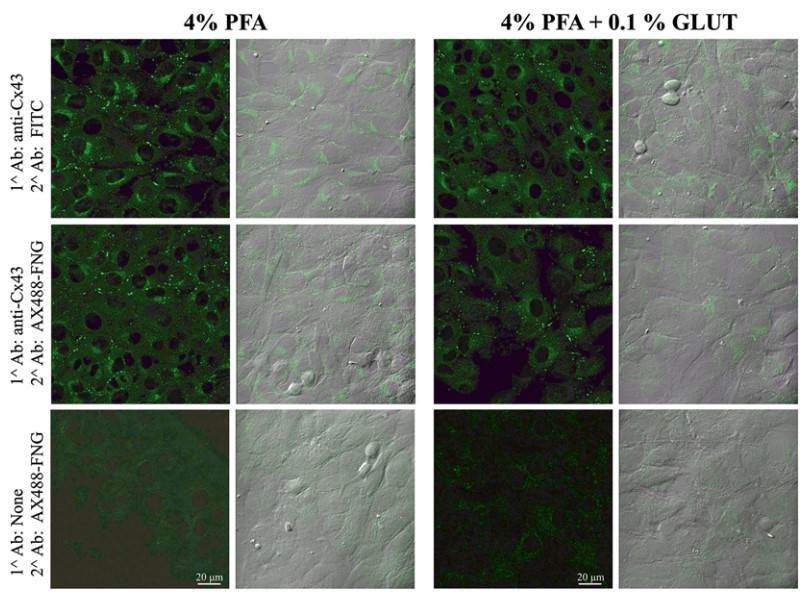 Figure 1. Immunostaining of endogenous Cx43 in NRK cells.
- Postfix with 2 % glutaraldehyde in PBS for 10 min at 4 °C.
- Remove fixative and rinse three times for 2 min each in icecold 1× PBS. Then wash cells for 5 min in quenching solution at 4 °C to remove aldehydes ( see Note 4).
- Rinse three times for 5 min each in ddH2O at 4 °C.
- Gold enhancement: incubate cells in the Gold Enhancement kit for 1–5 min to intensify gold particles. The reaction is light insensitive so it can be carried out under normal room lighting. The kit is composed of 4 components: the enhancer (A), the activator (B), the initiator (C), and the buffer (D). Equilibrate the solutions at room temperature. Right before use, combine equal volumes of A and B first, wait 5 min then add equal amounts of C and D ( see Note 5).
- Thoroughly rinse three times for 5 min each in ddH2O at 4 °C.
- Dehydration (ethanol series): dehydrate in increasing concentration of ethanol: 20, 50, 70, 90, and 2× 100 % ice-cold ethanol for 2 min each. Then rinse with 100 % ethanol at room temperature for 2 min.
- Infiltration and embedding:
- Remove 100 % ethanol and replace with a 50:50 mixture of low-temperature resin (LR White): ethanol 100 % for 20 min at room temperature.
- Remove the mixture and replace with 100 % LR White for 1 h. MatTek dish can be stored overnight in unpolymerized resin at 4 °C.
- The following day allow three more changes in 100 % of LR White within an hour.
- Cold-cure procedure: In a glass vial add 10 ml of LR White and one drop of accelerator. Mix well but avoid introducing any air to the solution and immediately add to cells. Cover with a piece of Aclar Embedding film, avoiding any air bubbles. Place MatTek dish inside an aluminum dish with ethanol on ice to keep the temperature low. Keep it on ice until the resin polymerizes (30 min to an hour). Store at 4 °C.
- At this point the cells can be imaged at the LM level: the signal of the fluorescent label is retained after the embedding procedure and the enhanced gold particles can be viewed by transmitted light.
- Sectioning: choose a region containing labeled cells and mount and trim the LR-White block. Cut ultrathin sections (70– 90 nm) and collect them on EM copper grids. Dry grids on filter paper and store them in a grid case at room temperature. Examine the ultrathin sections by TEM ( see Fig. 3).

 Loading ......
