
Inquiry Basket
 Loading ......
Cluster of Differentiation 2 (CD2) is a glycoprotein found on the surface of cells. It is a member of the immunoglobulin superfamily (IgSF). CD2 is a pan T-cell marker and NK cell marker, and it is involved in cell adhesion, co-stimulation, and immune synapse formation.
Browse all CD2 related products
CD2 is a type I single-pass transmembrane glycoprotein of around 50–58 kDa which is a member of the immunoglobulin superfamily. The protein structure of CD2 underlies many of its functions. The extracellular, ligand-interacting region of CD2 consists of two tandem immunoglobulin-like domains. The membrane-distal Ig-like domain is of the variable (V-set) region of antibodies and mediates binding to its major ligand CD58. The membrane-proximal Ig-like domain is of the constant (C2-set) region and is linked to the V-set domain by a flexible hinge region. It is thought to function in stabilizing the structural integrity and spatial conformation of the V-set domain. The intracellular region of CD2 has no intrinsic kinase activity. It is proline-rich and contains several protein interaction motifs, which are crucial in signal transduction.
 Figure 1. Model of CD2 binding to LFA3
(Source: Binder C, et al. 2020)
The major physiological ligand for CD2 is CD58 (LFA-3), which is also an immunoglobulin superfamily member, and is expressed on almost all cell types, including APCs, epithelial cells and erythrocytes. CD2-CD58 binding is a relatively low affinity but fast interaction. This type of "sticky" contact is essential for stabilizing transient communication between immune cells. Such trans-cellular interaction forms the physical basis for the formation of the immunological synapse, providing the necessary temporal and spatial conditions for subsequent high-affinity recognition events, such as those between the T-cell receptor (TCR) and peptide-MHC complexes.
The expression of CD2 exhibits marked cell-type specificity. CD2 is constitutively and stably expressed on almost all thymocytes throughout their lifespan and on mature T cells. For this reason, it has long been used as a definitive marker of T-cell lineage. During T-cell activation and differentiation, the expression level and conformation of CD2 can be dynamically regulated, leading to changes in its signaling capacity.
In NK cells, CD2 is involved in target recognition and adhesion but is also directly an activating receptor which mediates cytotoxicity and cytokine secretion. In other cells, like in some subsets of B-cell lymphoma or during certain stages of development, CD2 expression may also be observed but this is not generally the case. Detection of CD2 expression allows for accurate enumeration and functional analysis of lymphocyte subsets with particular importance in clinical immune function testing, diagnosis, and prognosis.
Detection of CD2 expression enables effective enumeration and functional assessment of lymphocyte subsets, which holds significant value in clinical immune function testing, disease diagnosis, and prognosis evaluation.
The CD2-CD58 interaction is the first event during immune cell-cell contact. At the initial stage of T cell–APC contact formation, large numbers of CD2-CD58 heterodimers accumulate at the contact area, forming a weak link between the two cells. This adhesion force plays an important role in increasing the scanning efficiency of the APC surface by the T-cell receptor (TCR) and the capture of cognate peptide-MHC (pMHC). After recognition by the TCR, the CD2-CD58 molecules, along with other adhesion molecules (LFA-1/ICAM-1) and signaling molecules, move and cluster at the outer rim of the IS. They contribute to the generation of peripheral supramolecular activation cluster (pSMAC), which stabilizes the synapse.
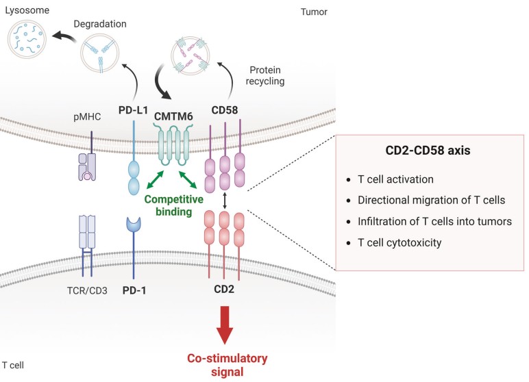 Figure 2. Regulation of tumor immunity and immune evasion via the CD2‒CD58 axis
(Source: Jo Y, et al. 2024)
CD2 serves as an important source of a "second signal" beyond the TCR signal. Upon TCR triggering (signal 1), binding of CD2 to its ligand CD58 on the target cell potently enhances the magnitude of TCR-induced downstream signaling intensity (co-stimulation). CD2 co-stimulation is essential for full T cell activation: it reduces the threshold for T cell activation, augments production of cytokines, particularly interleukin-2, leads to T cell proliferation and differentiation, and prevents the induction of anergy in T cells.
In NK cells, cross-linking of CD2 alone can directly trigger activating signals, inducing the release of perforin and granzymes to eliminate target cells.
Lacking any enzymatic activity in its cytoplasmic tail, CD2 nevertheless makes an efficient signal transducing platform. Cross-linking and clustering of the extracellular CD2 molecules bring the intracellular domain of CD2 into association with recruitment of signaling adaptor proteins and kinases and transduce downstream signals. CD2-associated protein (CD2AP) is one such adaptor that binds to the proline-rich sequence of CD2 cytoplasmic tail through its SH3 domain and transmits the signal to downstream effectors. A number of kinases including Lck and Fyn are found to associate with the cytoplasmic tail of CD2 and are activated upon cross-linking of CD2. Once activated, these kinases can phosphorylate the ITAM motifs of the TCR complex and other important signaling molecules (e.g. ZAP-70, LAT) thereby coupling the CD2 signal with the main TCR signaling pathway.
Furthermore, CD2 signaling can activate PLC-γ, leading to calcium influx and protein kinase C (PKC) activation, while also triggering pathways such as the MAPK cascade. These events ultimately lead to the activation of transcription factors (e.g., NF-κB, NFAT, AP-1), which regulate the expression of target genes.
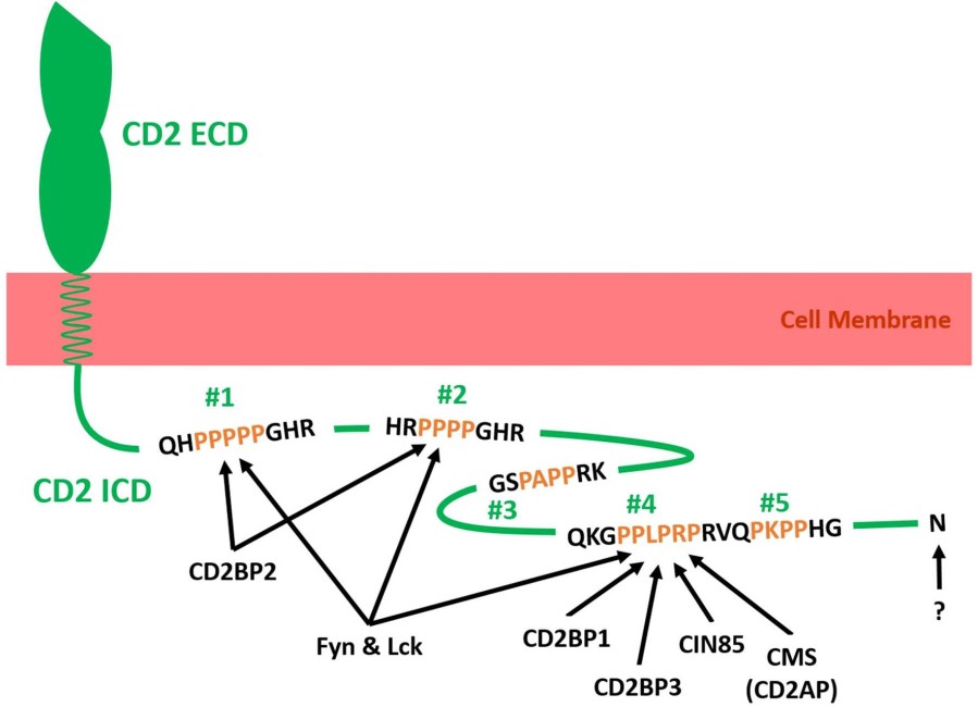 Figure 3. Schematic illustration of adaptor molecules binding to the intracellular tail domain of human CD2.
(Source: Binder C, et al. 2020)
CD2 has both positive and negative roles in the tumor microenvironment. For example, TILs with high CD2 expression are required for them to be anti-tumorigenic, and this anti-tumor activity of CD2 is through CD2-dependent adhesion/co-stimulation which allows the TILs to recognize and kill the CD58-expressing tumor cells. On the other hand, some tumor cells can escape T cell or NK cell killing by downregulating CD58 expression.
On the contrary, in some hematologic malignancies, for example, diffuse large B-cell lymphoma (DLBCL) or T-cell lymphoma, ectopic expression of CD2 or activation of the downstream signaling pathway may directly drive tumorigenesis and progression. In these situations, in addition to a biomarker for assessing the immune status of tumors, CD2 may also be a potential target for tumor immunotherapy in these particular malignancies.
Autoimmunity is when the immune system attacks the body's own tissues. Hyperactive CD2 co-stimulatory signalling may lower the activation threshold for autoreactive T cells, allowing them to be activated by low affinity self-antigens and thus promote or contribute to autoimmunity. Activated CD2+ T cell infiltrates have indeed been found at sites of inflammatory diseases such as psoriasis and rheumatoid arthritis and so inhibition of the CD2 pathway may be a potential treatment target for such conditions.
On the other hand, CD2-dependent signals are essential for efficient clearance of infectious pathogens such as virus and bacteria. The presence of defects in CD2 can result in immune defects and higher susceptibility to infections. In allergic disease, such as allergic asthma, CD2 is known to be associated with activation and function of Th2-type T cells, and plays a pathologic role in the allergic inflammation process.
References
Loading ......
