
Inquiry Basket
 Loading ......
When looked at closely, proteins, both intracellular and secreted, display a bewildering number of modifications. Because of the importance of posttranslational modifications for regulating biological activities of proteins there is a corresponding effort aiming at detecting and monitoring their occurrence. An increasing number of antibodies are becoming available that allow specific detection of modifications on selected proteins, mostly well-characterized components of signaling cascades. Such antibodies are, however, often not available for other proteins. The method described in this chapter can be used to detect and track protein modifications that lead to alterations of their charge and consequently of a change of the isoelectric point. Such charge isoforms can be separated by isoelectric focusing and are evident from two-dimensional separations where trains of spots are seen that are frequently due to different phosphorylation states. Especially high resolution is achieved by IEF on immobilized pH gradient gels. Although SDS-PAGE is sometimes capable of resolving modified proteins, the extent or even direction of the changed migration is difficult to predict.
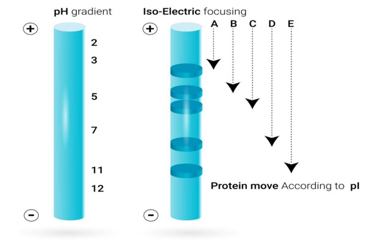 Figure1. IEF blotting.
The IEF blotting method described here was developed for monitoring alterations occurring on intracellular proteins that require denaturing conditions for extraction and separation. The method allows sensitive detection and estimation of the relative abundance of isoforms with antibodies. The latter is a quantity that is not easily measured by modification-specific reagents. Quantification is facilitated by running samples side by side under the same conditions. In principle, similar information can be obtained from two-dimensional separations by running a series of westerns. Results from such experiments are, however, difficult to quantify because of unavoidable technical variability. The separation method used here is very similar to the first dimension of two dimensional separation systems. It uses commercially available precast gels and allows direct comparison with two dimensional separations. Transfer from the plastic-supported gels is achieved by diffusion blotting.
Collect cells by centrifugation, wash once with PBS, suspend the pellet, transfer into a tared microcentrifuge tube, centrifuge once more, and remove as much of the supernate as possible. Estimate the volume by weighing, add R-buffer corresponding to five volumes of the cell pellet, suspend, and leave on ice for about 1 h. Centrifuge at 1,000 × g for 15 min in the cold. Store the sample at −80 °C. Protein concentration may be determined by a Coomassie binding assay. Protein concentrations are in the range of 3–10 mg/mL.
In order to assess any disturbances or differences between samples we find it useful to visualize and photograph the total protein pattern on the PVDF membrane using stains that are compatible with subsequent immunostaining. Also, if needed, IEF standards may be added in adjacent lanes and thus be visualized on the membrane.
It is useful to judge whether an assumed modification can account for the observed shift of the isoelectric point of the protein under investigation. Thus, for proteins with known amino acid composition, the isoelectric point can be predicted. The effect of phosphorylation can also be calculated. For acetylation on lysine groups, it is possible to simply omit one lysine from the sequence before computation of the pI. Similarly, other modifications may be incorporated by considering their effect on the charge of the protein.
Reference
Loading ......
