
Inquiry Basket
 Loading ......
Bacteria release membrane vesicles (MVs) with sizes ranging from 20 to 400 nm in diameter that affect diverse biological processes, including virulence, horizontal gene transfer, export of cellular metabolites, phage infection and cell-to-cell communication. Microbial membrane vesicles (MVs) also have immunomodulatory activities and are therefore used as vaccines and show great potential for the development of anticancer drugs and for applications in nanotechnology. Bacterial membrane vesicles (MVs) are abundant in coastal and open-ocean sea water, which implies that these structures are important for carbon cycling in the marine ecosystem. Membrane vesicles (MVs) are also abundant in microbial biofilms, where they are an integral constituent of the biofilm matrix and can protect biofilm cells from certain antibiotics.
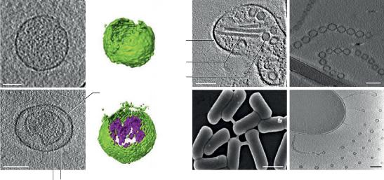 Fig1. Types of membrane vesicles. (Nature Reviews | Microbiology, 2019)
Outer-membrane vesicles. OMVs are the archetypal bacterial MV. They are spherical particles that consist of an outer leaflet of lipopolysaccharide (LPS) and an inner leaflet of phospholipid, which is derived from the outer membrane of Gram-negative bacteria. Classic OMVs originate from blebbing of the outer membrane and are therefore enriched for outer-membrane proteins, show specific lipid compositions and differ in the amount and content of cargo molecules depending on growth conditions. Many studies have shown that they contain periplasmatic and cytosolic proteins, DNA and RNA and that they transport virulence factors.
Outer-inner membrane vesicles. The first experimental evidence for MVs consisting of both the outer and the inner membranes was provided by a transmission electron cryomicroscopy study of Shewanella vesiculosa M7 supernatant, which unambiguously demonstrated the production of double bilayered MVs, which were originally named O-IMVs (Outer-inner membrane vesicles). Diverse bacteria produce OIMVs and that DNA is specifically packed into this type of MV.
Cytoplasmic membrane vesicles. Vesicle formation has been demonstrated in numerous Gram-positive bacteria. Given the lack of an outer membrane, we suggest that MVs produced by Gram-positive bacteria be named Cytoplasmic membrane vesicles (CMVs).
Tube-shaped membranous structures. Several different bacteria produce tube-shaped membranous structures (TSMSs), often referred to as nanotubes, nanowires or nanopods. These structures are tube-like protrusions of the cytoplasmic membrane of Gram-positive bacteria or of the outer membrane of Gram-negative bacteria and are considered to be specialized types of MVs. They often decorate the surface of the producing cell and form bridges between cells that enable the exchange of various cellular components.
Evidence has accumulated that suggests that two principal routes for the formation of membrane vesicles (MVs) exist: blebbing of membrane material of living cells that gives rise to classic outer membrane vesicles (OMVs), and endolysin-triggered cell lysis that leads to the formation of outer-inner membrane vesicles (OIMVs), explosive outer membrane vesicles (EOMVs) and cytoplasmic membrane vesicles (CMVs).
Treatment of bacteria with sublethal concentrations of certain antibiotics is a well-established trigger of bacterial vesicle formation. At least three mechanisms by which antibiotics stimulate membrane vesicle (MV) formation can be distinguished: cell envelope stress caused by antibiotics, induction of the SOS response and inhibition of cell wall biosynthesis. Antibiotics that weaken the cell wall, such as β-lactams, stimulate cytoplasmic membrane vesicle (CMV) formation in Gram-positive bacteria. This treatment will generate holes in the peptidoglycan layer through which cytoplasmic membrane material can protrude into the extracellular space and be released as CMVs. It has been well established that some antibiotics, particularly quinolones such as ciprofloxacin, induce the cellular SOS response, which in turn has been shown to stimulate vesicle production. As the SOS response triggers expression of endolysins encoded by prophages, these antibiotics stimulate vesicle formation through lysis. This pathway seems to be the main route for outer-inner membrane vesicle (OIMV) production. Antibiotics that cause cell envelope stress, such as polymyxin or gentamicin, will cause outer-membrane vesicle (OMV) formation through blebbing of the outer membrane.
Certain stimuli trigger specific outer-membrane vesicle formation routes. Evidence has accumulated that vesicle production not only depends on the genetic background of the producing strain but also is strongly influenced by the growth conditions. Factors triggering vesicle formation through blebbing. Forward genetic screens in several bacterial species have identified several genes that affect OMV production through blebbing. Factors triggering vesicle formation through endolysin-triggered cell death. Vesicle formation through explosive cell lysis or bubbling cell death depends on the expression of phage-derived endolysins.
Loading ......
