Introduction of Canine ELISPOT Assay
Vectors based on the adeno-associated virus (AAV) have shown promise for gene therapy of a number of acquired and inherited diseases in preclinical studies and clinical trials. However, one major hurdle in using viral vectors for in vivo gene delivery is the development of host cellular immune responses to the vector which may lead to elimination of transgene expression.
Cellular immune responses are mediated through the recognition of peptide epitopes presented by major histocompatibility complex (MHC) molecules on antigen-presenting cells by T-cell receptors, and can be assessed by several approaches. The chromium release assay has been a standard to measure specific cytotoxic T lymphocyte (CTL) activity. However, this assay requires knowledge of the peptide epitopes that are recognized and MHC molecule-matched target cells. The chromium release assay has low sensitivity and is not practical for assessing consecutive blood samples or for high-throughput screening to identify new epitopes. The use of a flow cytometry-based staining assay with MHC tetramers is more sensitive for detecting specific T cells than a chromium release assay, but depends on prior knowledge of both the MHC-restricting allele and the immunogenic epitopes. The other disadvantage is that MHC tetramer staining measures only the frequency of T cells with a specific T-cell receptor and not the function of the T cell. Intracellular cytokine staining (ICS) is another developed flow cytometry-based assay that can detect cytokine expression from responding T cells stimulated with peptides or peptide pools. It is a sensitive assay without the need of knowing the precise MHC-restricting allele, and when combined with multiparameter staining with antibodies to diverse cell surface markers can simultaneously detect the phenotypes of the responding T cells. However, the technique does not lend itself to high-throughput testing.
Interferon gamma (IFNγ) ELISPOT assay is a cytokine enzyme-linked immunospot assay that has many advantages over other methods. It can be used on blood samples without the need to add additional MHC molecule-matched target cells. IFNG ELISPOT can be designed in a high-throughput format to screen all potential epitopes in a given viral antigen using an overlapping peptide library, and thereby identify novel immunogenic epitopes. It is both sensitive and quantitative, and does not require large number of cells. Furthermore, the ELISPOT assay can be performed on frozen cells which would allow testing of multiple samples from different subjects or conditions or consecutive samples from the same subject within the same assay. It has been applied in analyzing peptide-specific immune responses in patients with various diseases, including vaccine trials for monitoring cell immune responses.
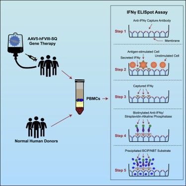 Figure 1. Monitoring cell-mediated immune responses using a validated IFN-γ ELISpot method. (Patton KS, et al.; 2021)
Here, we describe the use of INFG ELISPOT assay with a constructed panel of overlapping peptides spanning the entire VP1 sequence of AAV6 capsid protein to detect specific T-cell responses in peripheral blood in dogs given AAV6 vectors.
Methods of Canine ELISPOT Assay
Intramuscular AAV Vector Injection
All research experiments performed on dogs follow the Guide for Laboratory Animal Facilities and Care prepared by the National Academy of Sciences, National Research Council and after approval by the Institutional Animal Care and Use Committee. All dogs are immunized for leptospirosis, distemper, hepatitis, papillomavirus, and parvovirus and dewormed. Three muscles in hind limbs are chosen for intramuscular AAV injection for easy access: biceps femoris, semitendinosus, and semimembranosus.
- Shave injection sites.
- Dogs are under general anesthesia then placed in a lateral decubitus position; the site of incision is infiltrated with 2% lidocaine hydrochloride S.C.
- A 4–6-cm incision is made in the skin along the longitudinal axis of the hind limb to expose selected muscles.
- Nonabsorbable 4–0 sutures are placed in the belly of the muscle as marker for each injection site.
- Slowly inject 1 × 10¹¹ to 1 × 10¹² vector genome of rAAV vectors in 250 ML of HBSS per site into the muscle belly right underneath each suture using 31-gauge syringes.
- Close skin, and monitor animals daily for recovery.
Peripheral Blood Mononuclear Cells Collection
To examine cellular immune responses to AAV capsid proteins, blood samples are collected for isolation of PBMC before vector injection (pre) and at 4 and 12 weeks after AAV injection or at a desired time of your study.
- Collect 30 mL of blood containing 10% heparin as anticoagulant.
- Dilute blood in 30 mL prewarmed PBS (37°C).
- Transfer 15 mL of Ficoll into each 50-mL corning tubes, tilt the tube, and layer 30 mL of diluted blood very slowly on top of the Ficoll by gently pipetting the blood onto the side of the tube.
- Centrifuge at 365×g for 40 min at room temperature (RT) with the lowest speed acceleration rate.
- Remove the lymphocyte layer that collects above the Ficoll with a sterile plastic pipette, transfer to a 15-mL tube, and then fill the tube with Waymouth medium supplemented with 2% nonessential amino acid (Way-N) to 15 mL and centrifuge at room temperature for 10 min at 280×g.
- Remove supernatant, resuspend the cell pellet, and combine cells from all tubes and transfer to a new 15-ml tube. Fill the tube with Way-N and centrifuge at RT for 10 min at 224×g.
- Remove supernatant, and resuspend cells at 2 ×10⁶ cells/mL in 50% Waymouth, 50% ISCOVE medium supplemented with 1% nonessential amino acids, 1% sodium pyruvate, 1% penicillin streptomycin, 5% L-glutamine, and 10% HIDS (Way-ISC). Or alternatively, freeze down in 10% DMSO and 90% HIDS at a concentration of 10–15 × 106 cells/mL in liquid nitrogen.
ELISPOT Assay for Detecting T Cells to AAV Capsid Protein
To examine cellular immune responses to AAV capsid proteins, blood samples are collected at different time points described above for isolation of PBMC, and each sample is subjected for stimulation with peptides. PBMC collected before vector treatment stimulated with peptides and medium only and PBMC collected after vector treatment stimulated with medium only are used as negative controls. PHA stimulation is used as positive control for each sample.
Generation of Peptide Pools
- Obtain a peptide library consisting of 15-mer peptides each overlapping by 11 amino acids with adjacent peptides. HPLC purified peptides (purity >90%) are ideal.
- Dissolve each peptide in 100% DMSO at a concentration of 20 mg/mL. Number the peptide stocks sequentially and store at −80°C.
- Design a two-dimensional array for the individual peptides. For AAV6 VP1, there are 182 peptides in the panel, so a 14 × 13 array was designed.
- Generate a peptide pool by combining equal quantity of each peptide stock along an axis and a volume of DMSO sufficient to result in a final concentration of 2 mg/mL/peptide in each pool. For example, pool 1 contains all peptides in the first column, and pool 15 contains all the peptides in the top row. There are total 27 pools for AAV6 VP1. Store pools at −80°C until use.
ELISPOT Assay
- Add 50 ML of 70% ethanol to each well.
- Wash immediately 4× 200 ML of sterile PBS.
- Calculate the total volume of capture antibody needed and dilute to the working concentration using PBS.
- Add 100 ML diluted capture antibody to each well, cover, and incubate overnight at 4°C.
- Aspirate capture antibody and wash four times with wash buffer (0.05% Tween 20 in PBS, 350 ML/well). After the final wash, remove any remaining liquid by inverting the plate and blotting it against a clean paper towel.
- Block plates with 200 ML of blocking buffer (1% BSA, 5% sucrose in PBS) for 2 h at RT.
- During incubation, thaw peptide pool stocks and dilute each pool in culture medium (Way-ISC) to 4 Mg/mL. The culture medium is used as negative control, and 5 Mg/mL PHA is used as positive control.
- Aspirate blocking buffer, and wash the plates once with 350 ML of culture medium.
- Aspirate medium, and fill with 100 ML/well of each peptide pools or controls.
- Add 100 ML of PBMC suspensions (2 × 10⁶ cells/mL) to each well (final concentration of 2 × 10⁵ cells/well, see Notes 1 and 2) and incubate overnight at 37°C (see Note 3).
- Aspirate and wash plates four times with 350 ML wash buffer. After the final wash, remove any remaining liquid by inverting the plate and blotting it against a clean paper towel.
- Calculate the total volume of detection antibody needed and dilute to the working concentration using reagent diluent (1% BSA in PBS).
- Add 100 ML of the diluted detection antibody per well. Cover the plate with the lid and incubate overnight at 4°C.
- Aspirate and wash plates four times with 350 ML wash buffer. After the final wash, remove any remaining liquid by inverting the plate and blotting it against a clean paper towel.
Color Development
- Calculate total volume of streptavidin–AP needed and dilute 1:60 in diluent reagent.
- Add 100 ML to each well and incubate for 2 h at RT.
- Wash the plates 4× 350 ML wash buffer, rinse again with deionized water; after the final wash, remove any remaining liquid by inverting the plate and blotting it against a clean paper towel.
- Add 100 ML/well BCIP/NBT solution, cover, incubate for 30 min in dark at RT.
- Rinse six times with deionized water, invert plate, and tap to remove excess water.
- Allow plates to dry at RT for at least 3 h.
- Count spots using automated ELISPOT reader (see Notes 4 and 5).

 Loading ......
