
Inquiry Basket
 Loading ......
A variety of methods are available for the purification of antibodies, and the choice depends on the intended application, the species of origin, immunoglobulin (Ig) class and subclass, and the source of starting material. It is important to recognize that no single method can fulfill the requirements for all application needs, but those detailed in this chapter provide common methods suitable for most IgG purification. To maximize IgG purification, we take advantage of high-capacity IgG binding by specific bacterial proteins immobilized on a solid support such as agarose or Sepharose beads. The bacterial proteins used in the purification of antibodies are (1) protein- A derived from Staphylococcus aureus, (2) protein-G from Streptococcus sp., and (3) protein-L from Peptostreptococcus magnus. Each of these proteins provides unique Ig-binding properties, which can be used for purification of antibodies from a variety of matrices such as serum, culture supernatant, or ascites.
Polyclonal antibodies are frequently provided as an antiserum obtained from blood of immunized animals. Raw antiserum contains a large number of extraneous proteins that can interfere with immunoassays, and protein-A or protein-G is commonly used to eliminate the bulk of unwanted serum proteins. Antisera also contain a large number of unwanted antibodies (>95 % total IgG) against non-antigen targets, and these nonspecific Ig proteins are not removed by this purification method. Monoclonal antibodies are often provided as a hybridoma culture supernatant or as ascitic fluid harvested from the intraperitoneal cavity of mice after inoculation with a clonal hybridoma cell line. Similar to polyclonal antisera, monoclonal antibodies can be purified by protein-A or protein-G methods, but unlike antisera, the majority of immunoglobulins present in the crude fluid are of a singular class/subtype directed against the target antigen. Protein-L can often be substituted for protein-A or protein-G, and we take advantage of its inability to bind bovine IgG to purify mouse IgG from hybridoma culture supernatant, which is supplemented with fetal bovine serum (FBS). Neither protein-A nor protein-G bind IgY antibodies derived from chicken egg so purification is limited to protein- L. The typical concentration of antibody in antiserum is 1–3 mg/mL; hybridoma culture supernatant is 0.1–10 mg/mL and ascites 2–10 mg/mL. Prior to purification, crude antisera and ascites should be diluted in appropriate buffer to adjust binding pH and minimize bulk protein load effects that can block desired antibody binding to the resin.
Disposable column capable of containing at least 2 mL resin bed volume.
Immobilized protein-A, protein-G, or protein-L agarose/Sepharose beads (see Note 1).
Liquid chromatography system or handheld syringe-column system.
Antisera, ascites, or hybridoma culture supernatant.
Centrifuge.
Disposable centrifuge tubes (1.5–50 mL).
Disposable transfer pipette or syringe.
0.2μm filter.
1 N HCl.
1 N NaOH.
Thimerosal.
pH meter.
UV monitor for protein absorbance at 280 nm or protein assay such as Bradford or bicinchoninic acid (BCA).
Protein-A binding buffer: 0.1 M phosphate buffer, 0.15 M sodium chloride; pH 8.0.
Protein-A elution buffers: 0.1 M citric acid (pH 3.0–6.5).
pH 6.5 for IgG1.
pH 4.5 for IgG2a and IgG2b.
pH 3.0 for IgG3.
Protein-G binding buffer: sodium acetate (pH 5.0).
Protein-G and protein-L elution buffer: 0.1 M glycine-HCL (pH 2–3).
Protein-L binding buffer: 0.1 M phosphate buffer, 0.15 M sodium chloride; pH 7.2.
Neutralization buffer: high-ionic strength alkaline buffer such as 1 M phosphate or 1 M Tris (pH 7.5–9).
Dialysis bag/cassette or desalting column for buffer exchange.
Storage buffer: 30 % ethanol in water or 0.05 % thimerosal in water.
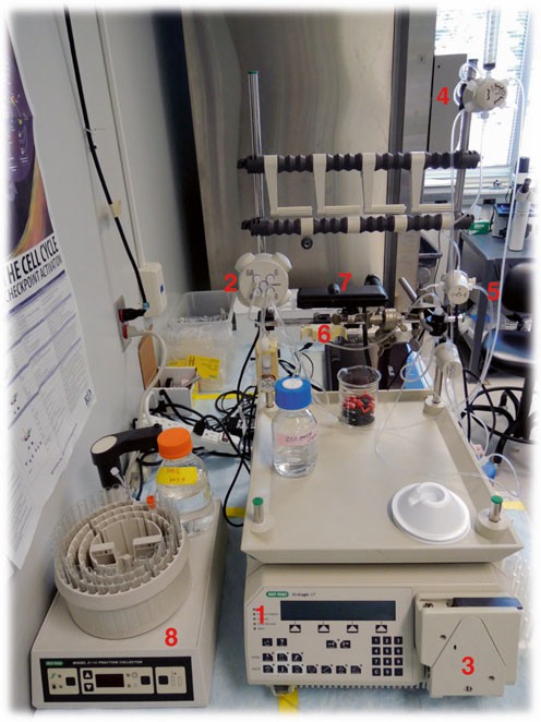 Figure. 1 An example of a liquid chromatography system with component parts and liquid plumbing.
Loading ......
