
Inquiry Basket
 Loading ......
As discussed in the previous chapter, a fresh tissue specimen received by the pathology laboratory must be fixed and dissected; fixation is most commonly achieved with formalin and dissection typically entails either complete or representative submission of tissue fragments into cassettes. Before microtomy, histochemical and immunohistochemical staining, and many other ancillary techniques can be performed, the cassetted formalin-fixed tissue must first undergo tissue processing to prepare the tissue for embedding within a supportive medium such as paraffin wax. Tissue processing is the subject of this chapter, and, in this introductory section, the theoretical underpinnings of the three sequential steps of tissue processing are described (dehydration, clearing, infiltration), followed by overviews of conventional tissue processing (both manual and automated) and microwave-assisted tissue processing.
The first step toward making aqueous formalin-fixed tissue fragments miscible with nonaqueous paraffin wax is dehydration, in which interstitial water is removed from the tissue. Historically, a variety of agents have been used to dehydrate tissues including alcohols such as ethanol, methanol, isopropanol, and butanol; acetone; dioxane; and tetrahydrofuran. Today, ethanol and isopropanol are probably the most commonly used dehydrating agents. These hydrophilic alcohols dehydrate tissues by drawing interstitial water out of the tissues by osmosis. Typically, the cassettes containing formalin-fixed tissue fragments are immersed in a series of alcohol baths of increasing alcohol concentrations. The alcohol concentration of the initial bath is usually in the 50–70% range; the alcohol concentrations in successive baths increase until an alcohol concentration of 100% is reached in the final bath(s). Using a graded alcohol series helps to ensure gradual removal of water thus mitigating damage to delicate tissues and undue tissue shrinkage that would occur if water were to be removed too abruptly, for instance by immediate immersion in 100% alcohol. Beginning at a low alcohol concentration also prevents the precipitation of phosphate salts from phosphate buffered formalin. It is essential that the concentration of the final alcohol in the graded series is 100% so as to promote complete removal of interstitial water from the tissues.
Following dehydration, the tissue is still not ready for paraffin infiltration because most dehydrating agents themselves are not miscible with paraffin. In the clearing step, the dehydrating agent is replaced by a clearing agent which is miscible with both the dehydrating agent and the paraffin. The clearing step also i) dissolves lipids, which is necessary as lipids and paraffin do not mix well, and ii) usually causes the tissue to become transparent due to the high index of refraction of most clearing agents. Many clearing agents used in the past (chloroform, benzene, dioxane, tetrahydrofuran, etc.) are toxic and/or carcinogenic and thus are unacceptable for present-day use. Xylene and toluene—in particular xylene—are now perhaps the most widely used clearing agents in contemporary histology laboratories. Clearing is accomplished by immersing the cassettes containing the formalin-fixed, now dehydrated tissue in several changes of xylene, for instance, to replace the dehydrating agent. Because xylene has its own associated health hazards and an unfavorable environmental profile, xylene-free clearing alternatives also exist and include D-limonene based products, alkanes/aliphatic hydrocarbons, alcoholic mineral oil mixtures, and, increasingly, the use of isopropanol as both a dehydrating agent and a clearing agent.
Once the dehydration and clearing sequence is complete, the tissue is suitable for infiltration with a supportive medium that will maintain the tissue's architecture during microtomy. Although a variety of media exist, paraffin wax is the most commonly used infiltration medium in histology and the discussion that follows is restricted to paraffin wax infiltration. Paraffin is produced from petroleum, and is a solid waxy substance at room temperature. Most commercially available paraffin preparations contain additives (latex, rubber, plasticizers, dimethyl sulfoxide, beeswax etc.) which influence various physical properties of the wax. Of these physical properties, melting point is particularly noteworthy. Very thin sections can be acquired using paraffins with higher melting points, and these relatively hard paraffins provide the appropriate level of support for hard tissues. However, paraffins with higher melting points do not form ribbons well during sectioning, and the heat required to melt them may introduce thermal artifacts and may inactivate antigens thereby interfering with immunohistochemistry. Furthermore, nucleic acid recovery is hampered by high temperatures. In contrast, while it is more difficult to achieve very thin sections from the softer more compressible paraffins with lower melting points, these paraffins generally ribbon well, are less susceptible to thermal artifact, and are better suited for subsequent immunohistochemical studies and nucleic acid extraction. Therefore, a laboratory must find the middle ground when choosing its paraffin preparation, and factors which influence this choice may include the properties of the tissue(s) processed by the laboratory, the volume of tissue being processed, and whether immunohistochemical or molecular studies are anticipated. Paraffin formulations for routine clinical use tend to have melting points in the 55–60 C range and it has been suggested that beeswax as an additive be avoided due to its potential to interfere with biomolecule recovery.
Infiltration is accomplished by immersing the cassettes of fixed, dehydrated, and cleared tissue in at least 2 changes of liquid paraf- fin. The paraffin infiltration step is incorporated into modern automated tissue processors, but otherwise should ideally occur in a vacuum oven. The incubation time in liquid paraffin should be long enough to obtain adequate infiltration, but not any longer than necessary in order to prevent prolonged exposure of the tissue to heat. For the same reason, it is recommended that the temperature of the liquid paraffin should be no more than 2–4 C above the paraffin's melting point.
The aforementioned steps of tissue processing can be accomplished in either a manual or an automated fashion. Manual processing is time-consuming, with the duration of most manual protocols exceeding the length of a regular work day. Furthermore, as compared to automated processing, manual processing is more susceptible to error and increases laboratory worker exposure to reagent fumes. Nevertheless, in resource-limited laboratories with low throughputs and/or in settings with an unreliable electrical supply, manual tissue processing may be necessary.
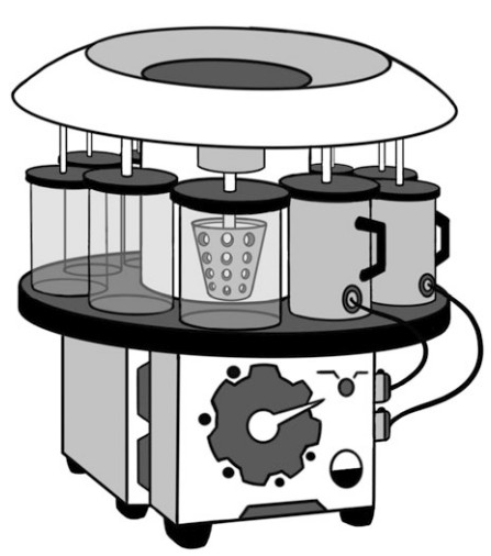 Figure 1. An automated tissue transfer/open processor transfers a basket containing tissues from one reagent bath to the next.
Conventional automated tissue processors come in two varieties: tissue transfer/open processors and fluid transfer/enclosed processors. Tissue transfer/open processors first became commercially available in the 1940s, and the earliest models had a rotary configuration with mechanical transfer of baskets containing tissue cassettes from one beaker of reagent to the next, thus circling through the steps of dehydration, clearing and infiltration. Given these design properties, tissue transfer/open processors are still sometimes referred to as "carousel" or "dip and dunk" processors. In addition to beneficial staffing and workflow implications related to the automated tissue changeover, these early processors also accelerated the speed of tissue processing by allowing for tissue suspension and agitation thus promoting more rapid reagent penetration. Modern tissue transfer/open processor models are computerized, and reagent exchange is further improved by the potential to apply vacuum and heat to any of the various processing steps.
The next major innovation in automated tissue processing was the development of fluid transfer/enclosed tissue processors. In these processors, baskets containing tissue cassettes are put into a chamber, referred to as a retort. According to a programmed schedule, the retort is then automatically and sequentially filled with and drained of the various processing reagents which are stored in a reagent cabinet incorporated into the instrument. Fluid transfer/enclosed processors are capable of higher throughputs than tissue transfer/open processors, and enhance the quality and speed of tissue processing by delivering agitation, vacuum/pressure, and heat (as appropriate) during processing thus improving reagent exchange. Fluid transfer/enclosed tissue processors also avert certain problems occasionally encountered with tissue transfer/open processors such as dropped tissue baskets or baskets hanging in the air and drying out. All modern fluid transfer/enclosed processors have fume control systems, and additional features may include multiple retorts, retort fill and pressure/vacuum level sensors, reagent monitoring, reagent autorotation, waste management systems, battery backup, remote monitoring systems in case of malfunction, and many others. Currently conventional fluid transfer/enclosed processors are widely used in high through put pathology laboratories, although certain practice settings now also use microwave-assisted tissue processing.
While most of the material in this chapter pertains to conventional tissue processing, an overview of microwave-assisted tissue processing follows. The concept of applying microwave energy to tissue processing is not new, but it is only in the last few decades that its use has become more mainstream. The basic physical premise of this technology is that microwave energy excites polar molecules within tissues (and reagents, if applicable) to generate instantaneous and uniform heat which, in turn, facilitates rapid reagent penetration. Fixation and the three subsequent processing steps of dehydration, clearing, and infiltration are all markedly accelerated by the application of microwave energy. Furthermore, microwave irradiation eliminates the need for a graded alcohol series to achieve dehydration, and microwave-assisted tissue processing is xylene-free because isopropanol is typically used for clearing. Altogether, microwave technology permits rapid tissue processing and can allow for same-day pathology reporting in clinical settings. Microwave processing has been shown to produce high quality morphological results, and most publications assert no negative impact on protein antigenicity or immunohistochemical labeling. A previous barrier to the implementation of microwave processing—that microwave use inserted manual steps into a process that was already automated—has been overcome now that high and continuous throughput automated microwave-assisted tissue processors are commercially available. Controversy remains as to whether accelerated formalin fixation achieved with microwave assistance is equivalent to time-based formalin fixation, in particular as it relates to performing prognostic/predictive ancillary tests; for instance, at the time of writing, guidelines still recommend a minimum formalin fixation time of 6 h for breast biomarker studies and do not distinguish between conventional or microwave-assisted formalin fixation and processing.
The reagent list provided is a general guide only; other reagent substitutes may be appropriate. Furthermore, the operating manual for the automated tissue processor being used should be always consulted to determine whether particular reagents are recommended or required.
Most of the reagents listed above pose human safety and/or environmental concerns. To mitigate risks, laboratory personnel must be appropriately trained to recognize the dangers of the materials used and take all appropriate precautions. Laboratory setup should also be optimized for safety. At a minimum, the following should be ensured.
The steps listed below are intended as a general guide only. Operating characteristics differ from one model of automated tissue processor to the next, and therefore the operating manual for the specific automated tissue processor being used should always be consulted.
Loading ......
