
Inquiry Basket
 Loading ......
Renal epithelial cells are actively involved in transporting water and solutes, as well as in homeostatic regulation. Trans-epithelial transport of substances is by either transcellular or paracellular pathways. Paracellular permeability is thought to be tightly associated with tight junctions (TJs). TJs are found in the most apical portion of the lateral membrane of epithelial cells and are made up of transmembrane proteins, cytoplasmic scaffold proteins, and signaling proteins. Transmembrane proteins, through their extracellular domains which extend into the intercellular space, help to regulate the movement of solutes via the paracellular route and are thought to be the primary determinants of paracellular permeability. Claudins are the most abundant transmembrane proteins in TJs.
Claudin family proteins have four transmembrane domains, and both N- and C-termini are on the intracellular side. This results in two extracellular loops of unequal size and a short intracellular loop. Claudins can be roughly divided into two major functional groups on the basis of their regulatory effect on permeability. The first group, best described as a "barrier", seals the paracellular pathway and limits free diffusion of molecules. Members of this group include Claudin-1, -3 and -5. The second group acts as selective "channels" through which some ions (usually cations or anions) can pass. Examples include Claudin-2, -4, -10, and -16.
Members of the Claudin family have characteristic expression and distribution patterns in different tissues and cells, as well as in different areas of the same tissue. Claudin genes are expressed in a segmental pattern in the kidney in specific nephron segments with each nephron segment expressing one or more Claudin proteins.
For instance, Claudin-2 is highly expressed in the proximal tubule. It is located mostly in the late part of proximal tubule and the early part of the descending thin limb of the loop of Henle. Claudin-2 forms a cation channel and mediates reabsorption of cations such as Na+ and K+. Claudin-4 and Claudin-8 are expressed in the distal tubule. Claudin-4 and Claudin-8 form a cation barrier that prevents the diffusion of Na+, K+ and H+ between the cells in the renal tubules. Claudin-4 and Claudin-8 thus help to maintain the cation concentration gradients needed for the transport of substances across the renal tubules (transcellular transport). In addition, Claudins can be alternatively spliced to produce Claudin-10 isoforms that have different extracellular domains and functional characteristics. Claudin-10a is anion selective and is expressed in the renal proximal tubule. Claudin-10b is very cation-selective, and is expressed in the medulla. Claudin-16 and -19 are found in the thick ascending limb of the loop of Henle where they mediate paracellular reabsorption of divalent cations and contribute to the positive luminal potential.
Claudins are not restricted to epithelial cells of the renal tubules; they have also been found in other parts of the kidney. For example, Claudin-7 is present in the connecting tubule and collecting duct. Claudin-9 is exclusively located in the proximal tubule, while Claudin-5 and Claudin-15 are primarily distributed in the renal vascular endothelium. Claudin-1 is found in renal glomerular epithelial cells, among others.
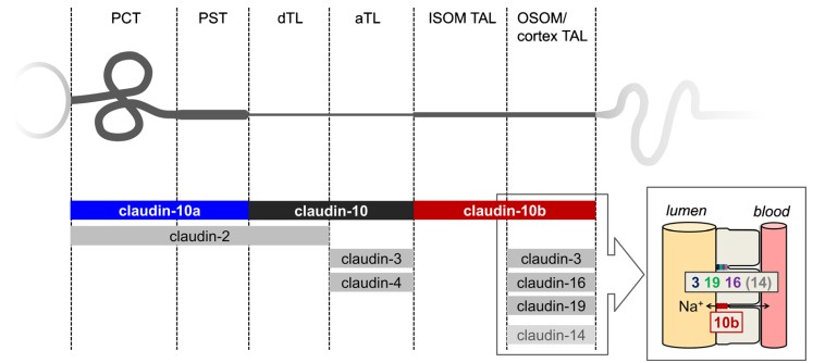 Figure 1. A schematic diagram of the distribution of certain Claudins in rodent nephrons
(Source: Milatz S, et al. 2017)
Parietal epithelial cells (PECs) are thought to restrict the escape of proteins filtered through the glomerulus to the periglomerular space. Tight junctions between PECs, together with the basement membrane at the cell base, constitute the second barrier for urine filtration. After experimental induction of glomerulonephritis, researchers found decreased Claudin-1 expression levels and increased permeability to albumin-sized tracers between adjacent PECs.
Claudins can selectively increase the permeability of cations or anions through the paracellular pathway. Claudin-4, -5, -8, -11 and -14 have been shown to selectively reduce cation permeability, particularly for Na+, K+, H+, and NH4+, while Claudin-2 and -15 have the opposite effect and increase cation permeability. Claudin-2 is mainly expressed in the proximal tubule and is essential for proper Na+ and water reabsorption in the proximal tubule and for the regulation of blood pressure. Claudin-2 gene knockout (KO) mouse models demonstrate significantly reduced net transepithelial absorption of Na+, C+, and water in the S2 segment of mouse proximal tubules.
Claudin-1 is positively expressed in both PECs and podocytes within the glomerulus starting from the S-shaped body stage of nephron development, but persists only in mature PECs. Podocytes are closely attached to the outer surface of glomerular capillaries, and the slit diaphragm (SD) between their secondary processes participates in forming the filtration barrier, serving as an important structure for primary urine formation. Using Claudin-1 transgenic mouse models, researchers induced Claudin-1 gene expression in mature podocytes, triggering severe proteinuria. Through freeze-fracture electron microscopy techniques, they discovered ultrastructural changes indicating SD-to-TJ conversion, confirming that Claudins and TJs play important roles in podocyte pathophysiology. The interaction between Claudins and SD components may promote SD-to-TJ conversion, suggesting that SD and TJ can be interchangeable under developmental and pathological conditions.
 Figure 2. Physiologic roles of Claudins in renal tubule epithelia
(Source: Yu AS. 2015)
The human Claudin-10 gene is located on chromosome 13q31-q34 and contains 5 exons. Within the first exon, there are 2 Claudin-10 splice variants that encode 2 major Claudin-10 isoforms: Claudin-10a and Claudin-10b. Research teams have described the clinical manifestations of biallelic mutations in the Claudin-10 gene, with the acronym HELIX (Hypohidrosis, Electrolyte disturbances, hypoLacrimia, Ichthyosis, Xerostomia) used to name this syndrome. All HELIX syndrome patients have functional defects in sweat glands, salivary glands, and lacrimal glands, with most patients exhibiting blood electrolyte abnormalities, hypermagnesemia and/or hypokalemia. Gene knockout experiments have shown that Claudin-10b specifically increases paracellular permeability to Na+. Indeed, the reduced paracellular permeability to Na+ in the thick ascending limb (TAL), sweat glands, and salivary glands may be the primary clinical consequence of loss-of-function mutations in Claudin-10b. In the renal medullary TAL, 50% of Na+ is passively reabsorbed through the paracellular pathway, and decreased paracellular permeability to Na+ affects Na+ reabsorption, leading to renal Na+ loss, extracellular fluid depletion, secondary aldosteronism, and renal K+ loss.
FHHNC is an autosomal recessive genetic disease caused by mutations in the Claudin-16 and Claudin-19 genes, characterized by massive loss of calcium and magnesium in urine, resulting in hypomagnesemia and hypercalciuria. Claudin-16 and Claudin-19 are expressed exclusively in the TAL and play crucial roles in paracellular transport of Ca2+ and Mg2+ in the TAL. When Claudin-16 is independently expressed in low-resistance LLC-PK1 cells, it can significantly increase paracellular Na+ permeability and moderately increase Mg2+ permeability, thereby changing selectivity from anion selectivity to slight cation selectivity. Expression of Claudin-19 in this cell line also eliminates anion selectivity, but in this case by reducing Cl- permeability. Co-expression of Claudin-16 and Claudin-19 results in more significant changes in Na+ and Cl- permeability than their individual expression levels, indicating functional synergy between these two Claudins.
Genome-wide association studies have confirmed that Claudin-14 is a major risk gene for hypercalciuric nephrolithiasis. According to genome-wide standards, synonymous mutations at the Claudin-14 gene locus are associated with increased kidney stone risk and decreased bone density. Homozygous patients with the common synonymous variant rs219780 (C) have an estimated 1.64-fold increased risk of developing kidney stones compared to non-carriers. Overexpression of Claudin-14 in various cell lines can reduce the Na+/Cl- permeability ratio as well as Ca2+ permeability. For example, in transfected LLC-PK1 cells, Claudin-14 can reduce the extracellular cation permeability of Claudin-16 through physical interaction.
Claudin proteins have been found to play important roles in cancer initiation and progression, and have shown great promise for cancer prevention and for fighting against cancer and its complications. Claudin-1 expression in squamous cell carcinoma was shown to be higher than in normal esophageal mucosa in one study, and Claudin-3 and Claudin-4 expression was found to be significantly higher in Barrett's esophagus and esophageal adenocarcinoma than in the pit epithelium in one study. Expression of Claudin-2 was significantly higher in esophageal adenocarcinoma than in Barrett's esophagus, and was found to be elevated in esophageal adenocarcinoma and its precancerous lesions in another study.
In gastric intestinal metaplasia, expression levels of Claudin-1, -3, -4, -5, -7, and -18 are elevated, and increased expression of these Claudin proteins is associated with gastric precancerous lesion status. As metaplasia progresses to dysplasia, the expression levels of these Claudins remain elevated, with Claudin-4 and -7 being most significant. The expression of Claudin-1, -3, -4, -5 and -7 is downregulated in diffuse gastric cancer. On the other hand, Claudin-18 expression is increased. Claudin-18.2 downregulation is more common in intestinal-type gastric cancer compared with diffuse-type gastric cancer, and thus, Claudin-18 may be expressed differently in various gastric cancer types.
In gene expression profiling studies, Claudin-1 was identified as the most important distinguishing gene between sessile serrated adenomas/polyps (SSA/P) and microvesicular hyperplastic polyps (MVHP). Inflammatory bowel disease patients have been confirmed to have higher risk of tumor transformation. Several studies have provided evidence that upregulation of several Claudin proteins promotes tumorigenesis in ulcerative colitis, including Claudin-1, -2, -3, and -4. Studies have reported that Claudin-1 upregulation in colitis is mediated by TNF-α, and it is speculated that the connection between TNF-α and Claudin-1 leads to increased resistance of colorectal cancer cells to anoikis. Controlled deletion of Claudin-7 can cause severe intestinal inflammation in mouse small intestine, indicating that Claudin-7 may exert tumor suppressive effects through anti-inflammatory actions.
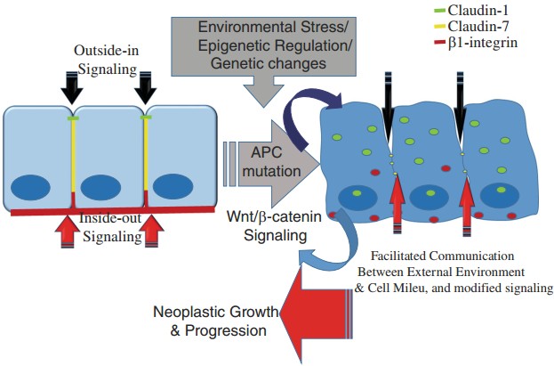 Figure 3. The regulation of Claudin-1 and Claudin-7 during colon tumorigenesis affects tight junctions
(Source: Singh AB, et al. 2017)
References
| Target | Cat. No. | Product Name | Size | Species | Application | Detection Sample | |
| CLDN1 | DEIA-XYA923 | Claudin 1 ELISA Kit | 96T | Qualitative | Cultured cells | Inquiry | |
| DEIA-LL259 | Rat Cldn1 (Claudin-1) ELISA Kit | 96T | Rat | Quantitative | Serum, plasma, tissue homogenates and other biological fluids | Inquiry | |
| CLDN11 | DEIA-XYA422 | Claudin 10 ELISA Kit | 96T | Qualitative | Cultured cells | Inquiry | |
| DEIA-XYA423 | Claudin 11 ELISA Kit | 96T | Qualitative | Cultured cells | Inquiry | ||
| CLDN2 | DEIA-FN286 | Human CLDN2 (Claudin-2) ELISA Kit | 96T | Quantitative | Serum, plasma, cell culture supernatants, tissue homogenate | Inquiry | |
| DEIA-FN287 | Porcine CLDN2 (Claudin-2) ELISA Kit | 96T | Quantitative | Serum, plasma, cell culture supernatants, tissue homogenate | Inquiry | ||
| CLDN4 | DEIA-XYA427 | Claudin 4 ELISA Kit | 96T | Qualitative | Cultured cells | Inquiry | |
| CLDN5 | DEIA-XYA428 | Claudin 5 ELISA Kit | 96T | Qualitative | Cultured cells | Inquiry | |
| DEIA-XYA429 | Claudin 5 (Phospho-Tyr217) ELISA Kit | 2 x 96T | Qualitative | Cultured cells | Inquiry | ||
| CLDN6 | DEIA-XYA430 | Claudin 6 ELISA Kit | 96T | Qualitative | Cultured cells | Inquiry | |
| CLDN7 | DEIA-XYA432 | Claudin 7 ELISA Kit | 96T | Qualitative | Cultured cells | Inquiry | |
| DEIA-XYA433 | Claudin 7 (Phospho-Tyr210) ELISA Kit | 2 x 96T | Qualitative | Cultured cells | Inquiry | ||
| CLDN9 | DEIA-FN288 | Human CLDN9 (Claudin 9) ELISA Kit | 96T | Quantitative | Serum, plasma, cell culture supernatants, tissue homogenate | Inquiry |
| Target | Cat. No. | Product Name | Host | Isotype | Application | |
| CLDN1 | CABT-L2897 | Mouse Anti-Human Claudin-1 monoclonal antibody, clone JID656 | Mouse | IgG | IHC | Inquiry |
| CLDN15 | CABT-L6363 | Rabbit Anti Human Claudin 15 polyclonal antibody | Rabbit | IgG | WB, ELISA | Inquiry |
| CABT-L6362 | Rabbit Anti Human Claudin 15 polyclonal antibody | Rabbit | IgG | IHC, IF/ICC | Inquiry | |
| CLDN18 | CABT-L1246 | Rabbit Anti-Human Claudin 18 monoclonal antibody, clone 45I25M26 | Rabbit | IgG | ICC, IHC-P, IF, WB | Inquiry |
| CABT-CS565 | Human Anti-Human Claudin 18.2 (Zolbetuximab) Monoclonal antibody, clone Zolbetuximab | Human | IgG1 | ELISA | Inquiry | |
| CLDN3 | CABT-L1247 | Rabbit Anti-Human Claudin 3 (phospho Thr192) monoclonal antibody, clone 5I28M39 | Rabbit | IgG | FC, ICC, IF | Inquiry |
| Target | Cat. No. | Product Name | Expression System | Tag/Conjugate | Application | |
| CLDN18 | DAG-WT1229 | Recombinant Human Claudin 18.2 VLP | HEK293 cells | N/A | ELISA, SPR | Inquiry |
| DAG-WT1230 | Recombinant Human Fluorescent Claudin18.2 VLP | Expi293 | GFP | ELISA, SPR | Inquiry | |
| CLDN6 | DAG-WT1223 | Recombinant Human Claudin 6 VLP | HEK293 cells | N/A | ELISA, SPR | Inquiry |
| DAG-WT1224 | Recombinant Cynomolgus Claudin 6 VLP | HEK293 cells | N/A | ELISA, SPR | Inquiry | |
| DAG-WT1225 | Recombinant Mouse Claudin 6 VLP | HEK293 cells | N/A | ELISA, SPR | Inquiry | |
| DAG-WT1244 | Biotinylated Recombinant Human Claudin 6 VLP | HEK293 cells | N/A | ELISA, SPR | Inquiry |
Loading ......
