Introduction of ELISPOT and ELISA to Single Cells' Cytokine
Although the enzyme-linked immunospot (ELISPOT) assay is thought to be more sensitive than ELISA, allowing for the detection of a single cytokine-secreting cell out of a million, the major limitation of the ELISPOT assay is that, unlike ELISA, it does not quantitate the amount of secreted cytokines. The output of an ELISPOT assay is a formation of colored spots which appear at the sites of cells releasing cytokines, and it appears that each individual spot represents a "footprint" of a single cytokine releasing cell.
ELISPOT allows detection of just a few cytokine-releasing cells out of tens of thousands, making this technique a method of choice for vaccine development, cancer research, AIDS research, allergy research, and autoimmune disease studies.
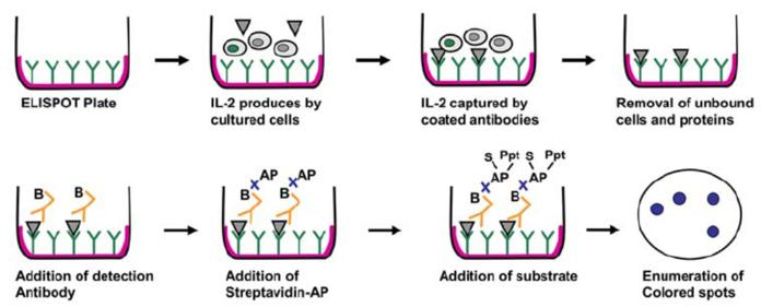 Figure 1. Detection of cytokines (eg: IL-2) released by cells using ELISPOT technique. (Naresh Sachdeva, et al.; 2007)
We worked out a simple protocol for the determination of the average amount of IFN gamma secreted by a single cell. Our protocol can be easily adapted for measuring other cytokines in different species and utilized for both basic research and for developing diagnostic applications.
Methods of ELISPOT and ELISA to Single Cells' Cytokine
Isolation of Human PBMCs
- Collect blood samples from healthy donors in standard citrate-phosphate-dextrose unit bags.
- Separate PBMCs using density centrifugation (500 × g for 30 min) by layering 25 mL of blood on 20 mL of 1.077 g/mL Ficoll-Paque Plus at 25°C (see Note 3).
- Discard the upper plasma layer after centrifugation and transfer the blood cells into two sterile 50-mL tubes.
- Resuspend the blood cells in 45 mL of sterile PBS and centrifuge for 5 min at 500 × g.
- Discard the supernatant, and resuspend the pellet in 10 mL of red blood cell lysing solution and incubate for 5 min at room temperature.
- After lysing, resuspend PBMCs by adding sterile PBS up to the 50-mL graduation mark on the tube.
- Centrifuge the tubes for 5 min at 500 × g.
- Discard supernatants and add 30–40 mL of RPMI complete medium to the tubes with PBMCs.
- Mix cells 1:2 with Trypan blue dye and pipette 10 μL of that mixture into each side of a hemacytometer under a coverslip (see Note 4). Count the cells under the microscope using a 20× lens and a phase-contrast condenser.
- Isolated PBMCs are divided into two pools and cells from one pool are cultured in a regular ELISPOT plate, whereas cells from the other pool are cultured in an uncoated, blank, ELISPOT plate.
ELISPOT Assay
Cells are plated into both coated ELISPOT plates and plates not coated with capture anti-IFN gamma antibodies.
- Prepare uncoated ELISPOT plates and culture PBMCs as follows:
- Block ELISPOT plates by adding 100 ML of block buffer per well and incubate for 1.5–5 h at room temperature.
- Discard block buffer and fill each well with 200 ML of RPMI complete culture media. Allow to sit a minimum of 20 min and discard the culture media just before plating PBMCs.
- Plate PBMCs (100 μL/well; six wells per group) into both uncoated and coated ELISPOT plates at cell concentrations of 104 and 105 cells/mL (see Notes 5 and 6).
- Stimulate PBMCs with a mixture of CaI and PMA by adding them directly to the ELISPOT plate, so their final concentrations reach 0.5 Mg/mL and 50 ng/mL, respectively.
- Incubate in a CO2 incubator at 37°C for 18 h (see Notes 7 and 8).
- After incubation, collect the supernatant mixture with a multichannel pipette from both uncoated and coated plates for subsequent ELISA analysis. (Uncoated plates can be discarded at this time, but coated plates are processed to develop spots.) Then, wash the plate by rinsing wells four times with wash buffer to remove PBMCs from the plate (see Notes 9 and 10).
- Prepare working solutions of detection antibodies by mixing concentrated detection antibodies 1:120 with dilution buffer.
- Add 100 μL of detection antibody working solution into each well and incubate ELISPOT plates overnight at 2–8°C.
- Wash plates three times with the wash buffer.
- Prepare a working solution of streptavidin–alkaline phosphatase by mixing the concentrated stock solution 1:120 with corre-sponding dilution buffer.
- Add 100 μL of streptavidin–alkaline phosphatase working solution into each well and incubate for 2 h at room temperature.
- Wash plates three times with wash buffer.
- Add 100 μL of ready-to-use BCIP/NBT substrate into each well and incubate for 30–60 min at room temperature in a place protected from direct light.
- Wash plates three times with distilled water and let them dry completely (see Note 11).
- Quantify spots using an automated ELISPOT reader.
ELISA Assay
- To remove contaminating PBMCs, centrifuge supernatants collected from coated and uncoated ELISPOT plates at 500 × g.
- Pipette 500 mL of Calibrator Diluent RD6-21 into polypropylene tubes. Use the kit standards stock solution to prepare a dilution series. Mix each tube thoroughly before the next transfer. The undiluted standard serves as the high standard (1,000 pg/mL). Calibrator Diluent RD6-21 serves as the zero standard (0 pg/mL).
- Add 100 μL of Assay Diluent RD1-51 to each well. Add 100 μL of standard or sample to each well (see Note 12). Cover with an adhesive strip and incubate for 2 h at room temperature.
- Aspirate and wash the plate four times with wash buffer (see Note 10).
- Add 200 μL of conjugate to each well. Cover with an adhesive strip and incubate for 2 h at room temperature.
- Aspirate and wash the plate four times with wash buffer (see Note 10).
- Add 200 μL of substrate solution to each well. Incubate in the dark for 30 min at room temperature.
- Add 50 μL of stop solution to each well. The color should change from blue to yellow (see Note 13).
- Determine the optical density of each well within 30 min using a microplate reader set to 450 nm. If wavelength correction is available, set to 540 or 570 nm (see Note 14). If wavelength correction is not available, subtract readings at 540 or 570 nm from the readings at 450 nm. This subtraction corrects for optical imperfections in the plate. Readings made directly at 450 nm without correction may be higher and less accurate.
Calculation of the Amount of Cytokine per Spot (See Note 15)
- Calculate the average amount of IFN gamma in the culture media collected from the uncoated plate and from the coated ELISPOT plate measured by ELISA.
- Calculate the average number of spots counted with an ELISPOT reader.
- Subtract the average amount of unbound (UB) IFN gamma from the total (T) amount of IFN gamma per well (0.1mL) and divide it by the average number of spots (N) to calculate the average amount of IFN gamma per each spot (C spot):

This simple protocol allows for the calculation of the average amount of IFN gamma secreted by a single cell that can be easily adapted for measuring other cytokines in a variety of species.
Although this approach has some limitations, it can be quite useful for estimating the extent of cytokine production at a single-cell level. This protocol can be easily adapted for measuring other cytokines and used for both a basic research as well as for developing diagnostic applications.

 Loading ......
