
Inquiry Basket
 Loading ......
Rhabdomyosarcoma is a malignant tumor that originates from striated muscle cells or mesenchymal cells that differentiate into striated muscle cells. It is the most common type of soft tissue sarcoma in children. The incidence rate of rhabdomyosarcoma is second to malignant fibrous histiocytoma and liposarcoma, ranking third in soft tissue sarcoma. Adults have less hair, with more males than females. Embryonal rhabdomyosarcoma is more common in children before the age of 8 (with an average age of 6 years); Alveolar rhabdomyosarcoma is common in adolescent males (with an average age of 12 years); Multi type rhabdomyosarcoma is common in adults and can also be seen in children.
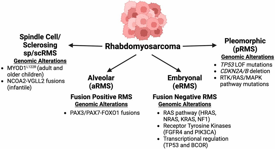 Figure 1.Rhabdomyosarcoma subtypes with prominent genomic alterations.( Gustafson, Annika L,et al, 2025)
Embryonal rhabdomyosarcoma accounts for about 2/3 of rhabdomyosarcoma, and is more common in children and adolescents. Commonly found in the head, neck, urogenital tract, and retroperitoneum. The disease course is short, and the main symptoms are painful or painless lumps, redness and swelling on the skin surface, and high skin temperature. The tumors vary in size, are hard in texture, and most of them are fixed when seeking medical attention. Tumors grow rapidly and may cause skin rupture and bleeding. Pain may occur when tumors compress nerves. Head and neck lumps may have eyeball protrusion, bloody discharge, nosebleeds, swallowing and respiratory disorders. Urogenital system tumors are characterized by vaginal bloody secretions, hematuria and urinary retention, and pelvic masses can be touched by anal fingers. This type often metastasizes to retroperitoneal lymph nodes and regional lymph nodes, and in late stages, it is often accompanied by hematogenous metastasis.
HE staining is used to observe rhabdomyosarcoma cells under an optical microscope, which can sometimes be difficult to diagnose due to low cell differentiation and absence of striations. Immunohistochemistry staining is a reliable method for diagnosing rhabdomyosarcoma. Under an optical microscope, the tumor cells in this case were observed to be arranged in a bundle shape, resembling a fibrosarcoma. A few tumor cells were found to have obvious eosinophils in the cytoplasm, namely rhabdomyoblasts. Immunohistochemistry showed positive Myoglobin. Generally, there are no typical radiological features or calcification phenomena. Tumors can invade and destroy adjacent bone, especially in the skull, forearm, hands, and feet. CT and MRI examinations with contrast agents can effectively display the location, volume, edges, and relationship with surrounding tissues of tumors. Intravenous pyelography can detect irregular filling defects and hydronephrosis in the bladder. Other examination methods include bone scan (suspected of bone metastasis) and lymphangiography (suspected of lymph node metastasis)
| Diagnosis | Description |
| Head | Head and neck rhabdomyosarcoma is mostly embryonal in children, while it is grape shaped sarcoma in the ears, nose, and sinuses. The patients presented with lumps, which include symptoms such as protrusion of the eyeball, changes in voice, difficulty swallowing, respiratory obstruction, cough, and discharge from the external auditory canal. If it invades the nerves, acid reflux pain will occur; The swelling and invasive growth of tumors can worsen symptoms and lead to the appearance of brain symptoms. If there is a transfer, corresponding symptoms will occur. |
| Eye sockets | Rhabdoblastoma can come from the eye muscles or lacrimal glands, and is more common in boys aged 7-8 years, causing unilateral exophthalmos. The condition progressed rapidly, with one-third of the patients experiencing upper eyelid ptosis, 10% experiencing headaches, and X-ray showing bone destruction. If a child has unilateral protruding eyes, neurological examination, including carotid angiography and brain scan, should be performed. Differential diagnosis should include leukemia and neuroblastoma. Leukemia can be easily distinguished by peripheral blood count and bone marrow smear. When neuroblastoma invades the orbit, the lesions in other parts of the body are more obvious and can be distinguished. |
| Ear | Tumors can occur in the external auditory canal, middle ear, mastoid process, or paranasal sinuses, often affecting only one ear and normal contralateral side. They often have unilateral hearing loss and often go unnoticed. Often diagnosed with polypoid masses in the external auditory canal and bloody secretions in the ear, it is easy to mistake them for inflammatory polyps. Therefore, when young children have ear inflammation that is ineffective with antibiotic treatment, this disease should be considered in particular. Less common is the mass on the outer side behind the ear, which grows rapidly. At the initial diagnosis, it may be an inconspicuous protrusion with a soft texture. At follow-up, it can enlarge to an alarming degree. Due to its painlessness, the diagnosis is often in the late stage. Occasionally, the chief complaint is facial nerve paralysis, and dizziness is a relatively late stage symptom. The tumor spreads from the middle ear to the mastoid process and invades the posterior cranial fossa through the inner plate. |
| Mouth and neck | Rhabdomyosarcoma is divided into embryonal, acinar, and polymorphic types, with 80% of rhabdomyosarcoma occurring in the orbit being embryonal. Embryonal rhabdomyosarcoma includes spindle cell, grape shaped, and anaplastic rhabdomyosarcoma, which are easily confused with tumors such as non Hodgkin's lymphoma and fibrosarcoma. |
1. Surgical treatment
Surgical resection is the main method for rhabdomyosarcoma, and the resection range includes all muscles where the tumor is located. For embryonal rhabdomyosarcoma, in addition to resection, chemotherapy and radiotherapy should also be combined to alleviate symptoms; Polymorphous rhabdomyosarcoma is ineffective in chemotherapy and radiotherapy treatment. The biopsy of the tumor showed that complete surgical resection of rhabdomyosarcoma resulted in better outcomes. Only 10% of patients' rhabdomyosarcoma can be completely removed. Even for patients who have undergone complete resection, chemotherapy and radiotherapy are necessary because rhabdomyosarcoma is highly prone to metastasis.
2. Chemotherapy
When rhabdomyosarcoma cannot be completely removed, patients must undergo chemotherapy. Chemotherapy can completely eliminate residual tumors. Even if the tumor appears to have been completely removed, chemotherapy is still necessary.
3. Radiotherapy
For rhabdomyosarcoma, radiotherapy is a very effective method and can be used as an adjuvant therapy for surgical treatment. The radiation dose should be selected according to age and location, and the radiation field should include the tumor bed and surrounding normal tissue of 2-5 centimeters. The effective radiation dose should not be less than 40Gy.
4. Magnetic induction therapy
Magnetic induction therapy utilizes the principle of ferromagnetic materials generating heat under an alternating magnetic field to raise the temperature of tumor tissue to an effective level, thereby achieving the therapeutic goal. Magnetic induction therapy has the characteristics of targeting, conformity, self controlled temperature, internal heating, repeatability, and large temperature difference between normal tissue and tumor. It is expected to overcome the shortcomings of previous local hyperthermia methods for tumors and become a new effective means of treating tumors.
The location of onset of rhabdomyosarcoma will affect the prognosis. Patients who occur in the head, neck, and urogenital areas have a better prognosis, while those who occur in the limbs and trunk have a poorer prognosis. At present, the comprehensive treatment of surgery, radiotherapy, and chemotherapy, if there is no metastasis before starting treatment, has a 5-year survival rate of nearly 80%. Two thirds of children with rhabdomyosarcoma can survive, and the most important factor is the tumor resection: children with type I rhabdomyosarcoma have good treatment outcomes, with over 90% of them not relapsing; 80% of Class II and 70% of Class III will survive for a long time; The prospects for Class IV children are not good, with a 5-year survival rate of less than 30%.
Reference
| Cat. No. | Product Name | Size | Species Reactivity | Application | Detection Method | |
| DEIA-XYA688 | FOXO1 ELISA Kit | 96T | Human,Mouse,Rat | Qualitative | iELISA | Inquiry |
| Cat. No. | Product Name | Size | Target | Species | |
| DAG-KO191 | FOXO3 Knockout Cell Lysate | 100 g | / | / | Inquiry |
| CDBP3213 | Human XAGE1E blocking peptide | 100 g | XAGE1 (Isoform 1d) / GAGED2 | Human | Inquiry |
| CDBP3214 | Human XAGE1E blocking peptide | 100 g | XAGE1 (Isoforms 1a, 1b and 1c) | Human | Inquiry |
| CDBP5455 | FOXO1 blocking peptide | 0.05 mg | / | / | Inquiry |
| CDBP6202 | STIM1 blocking peptide | 0.05 mg | / | / | Inquiry |
| DAG-P1503 | Human FOXO3 (phospho S253) blocking peptide | / | / | / | Inquiry |
| DAG-P1504 | Human FOXO3 blocking peptide | / | / | / | Inquiry |
Loading ......
