
Inquiry Basket
 Loading ......
Poliomyelitis (Polio) is a highly infectious disease caused by the poliovirus. Since the inception of the Global Polio Eradication Initiative (GPEI) in 1988, the overall reduction in global incidence of poliovirus transmission has been more than 99% with the successful use of polio vaccines over the past several decades and effective programmatic use. More than 2.5 billion children have been immunized against polio, leading to its elimination from much of the world.
Poliovirus invades the nervous system and can cause total paralysis within hours. The virus is transmitted by person-to-person spread primarily through the faecal-oral route or, less commonly, by a common vehicle (for example, contaminated water or food) and multiplies in the intestine. The initial symptoms of poliovirus infection include fever, fatigue, headache, vomiting, stiffness of the neck, and pain in the limbs. One in 200 infections leads to irreversible paralysis, usually in the legs). Among those paralyzed, 5-10% may die if their breathing muscles become immobilized. It is important to note that polio can affect anyone of any age, although it predominantly affects young children under 5 years of age. Vaccination is key in preventing the spread of poliovirus and protecting individuals from the disease.
Poliovirus is a member of the genus Enterovirus, belonging to the Picornaviridae family. This virus has a positive-sense, single-stranded RNA genome enclosed within a non-enveloped ~30 nm protein capsid. The major open reading frame (ORF) is translated as a single polyprotein comprising regions P1 (encoding the viral capsid proteins) and P2 and P3 (proteins for proteolytic processing and replication). The viral protease precursor 3CD cleaves P15 into the capsid proteins VP0, VP1 and VP3, and encapsidation of the viral RNA to form the mature virion is associated with cleavage of VP0 into VP2 and VP4, increasing particle stability. The icosahedral protein capsid is composed of 60 protomers each made of 4 virion proteins: VP1, VP2, VP3, and VP4. All the 4 virions are made of 8 strands of protein arranged in β sheet array forming a β barrel. Due to the intermingling of various proteins, loops are created, which serve as antigenic sites for combination with corresponding antibodies. Three serotypes of poliovirus have been recognized as types 1, 2, and 3. The prototype strains are Brunhilde and Mahoney strains for type 1, Lansing and MEFI for type 2, and Leon and Saukett for type 3.
 Fig. 1 Schematic representation of the poliovirus genome (Bahar, M.W. et al. 2021)
Poliomyelitis is a human disease that is exclusively transmitted from an infected individual or a carrier without symptoms through the fecal-oral route. The spread is rapid in areas with poor sanitation, especially among the non-immune population. The propagation of the virus is mainly seen in summer months in temperate regions. Poliomyelitis has been endemic in susceptible infants through infection. When the poliovirus binds to the cell surface, the virus is internalized through a clathrin-, caveolin-, and flotillin-independent, but actin- and tyrosine kinase-dependent, pathway. After internalization, the virus quickly releases its RNA from vesicles that are located within 100-200 nm of the plasma membrane. This process does not require endocytic acidification or microtubule-dependent transport.
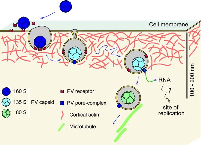 Fig. 2 Model of poliovirus entry (Brandenburg, B, et al. 2007)
Poliovirus can be detected in specimens from the throat, feces (stool), and occasionally cerebrospinal fluid (CSF) by isolating the virus in cell culture or by detecting the virus by polymerase chain reaction (PCR).
Serology may be helpful in supporting the diagnosis of paralytic poliomyelitis, particularly if a patient is known or suspected to not be vaccinated. When poliovirus serology is indicated, a pre-infection and post-infection serum is required, with a minimum volume of 0.5ml of each serum.
There are two vaccines available: live attenuated oral poliovirus vaccine (OPV) and inactivated polio vaccine (IPV). Both IPV and OPV were developed in the 1950s and have since been used worldwide for routine childhood immunization and to prevent and control polio outbreaks in endemic countries.
OPV is known to replicate efficiently in the intestine but is around 10,000 times less able to enter the central nervous system than the wild polio virus (WPV). Administration of OPV mimics the immune response to natural exposure to WPV generating both humoral and mucosal immunity. IgM antibody becomes detectable as early as 2-3 days after infection, usually disappearing after 2-3 months, while IgG becomes the predominate antibody and may last for life. There are different types of OPV, which may contain one, a combination of two, or all three different serotypes of attenuated vaccine. The type 2 strain of OPV is immunodominant, and most formulations of trivalent oral poliovirus vaccine (tOPV) have a 10:1:6 ratio of virus for the three serotypes, respectively. In the USA, after a complete primary vaccination series of three doses, over 95% of recipients seroconvert to all three poliovirus serotypes with long-lasting immunity.
The advantages of OPV include high levels of intestinal immunity, which prevent virus shedding and contribute to population protection. Additionally, OPV is easy to administer and is cost-effective, making it a suitable choice for resource-constrained, high disease burden settings. However, OPV can rarely cause vaccine-associated paralytic poliomyelitis (VAPP) in vaccine recipients and close contacts. Furthermore, the live vaccine virus has the potential to mutate into forms that resemble wild polioviruses, leading to outbreaks known as circulating vaccine-derived polioviruses (cVDPVs).
IPV is composed of inactivated (killed) strains of all three poliovirus types. Based on the data from the dose ranging studies and as a compromise between protective immune response in children and the quantity of vaccine that could be optimally manufactured with cost considerations, WHO defined in 1981 the 40-8-32 D-Ag units composition as the specification for the antigenic content for all trivalent IPV formulations. Since 2000, IPV is the only poliovirus vaccine available for routine infant and childhood immunization in the United States. It is administered through either intramuscular or intradermal injection. The current formulation of IPV induces close to 100% seroconversion rates with high antibody titers to the three poliovirus serotypes after a series of three doses, when administered in schedules in which the last dose is administered at 6 months of age or older. In most middle- and upper-income countries, IPV is the preferred vaccine for poliomyelitis. This is because IPV is considered safe, highly effective, and does not cause VAPP. Additionally, IPV can be combined with other vaccines and administered through intramuscular injection. Combination vaccines such as DTaP/IPV/Hib, DTaP/Hep B/IPV, and DTaP/IPV often include IPV as one of the components.
Even though there are highly effective vaccines to control poliomyelitis, there are still some challenges to overcome. Such as the occurrence of revertant neurovirulent strains of polioviruses from OPV, and the lack of primary intestinal mucosal protection from IPV. In the post-eradication period, the ideal vaccine would possess several characteristics. In the post-eradication period, the ideal vaccine would possess several characteristics. These include high humoral and intestinal immunity, long-lasting protection, low cost, ease of administration, safe production on a large scale, minimal waste, and heat and freeze stability. Efforts are being made to develop a vaccine that fulfills these criteria.
References
Loading ......
