Medica 2026
Nov 16-19, 2026 - Düsseldorf, Germany

Recombinant HPV 16 and 18 Antigens

Human papillomavirus (HPV) is a virus from the papillomavirus family that is capable of infecting humans. There are nearly 200 different strains of HPV, most of which are harmless and not cancer causing. While in those disease strains, clinical statistics shows that 14 high-risk HPV types (HPV 16, 18, 31, 33, 35, 39, 45, 51, 52, 56, 58, 59, 66, and 68) are known to lead to cancers of the cervix, vulva, vagina, penis, oropharynx and anus. Among them, two HPV types (16 and 18) cause 70% of cervical cancers and pre-cancerous cervical lesions. HPV 16 and 18 are more carcinogenic than other HPV types. With years of exploration, Creative Diagnostics has successfully obtained various highly purified HPV 16 and 18 antigens. Our products have significantly contributed to the HPV vaccine related research.

Structure of HPV

HPV is a relatively small, non-enveloped, epitheliotropic, double-stranded DNA viruses. The genomes of HPV contain three functional parts ORFs.
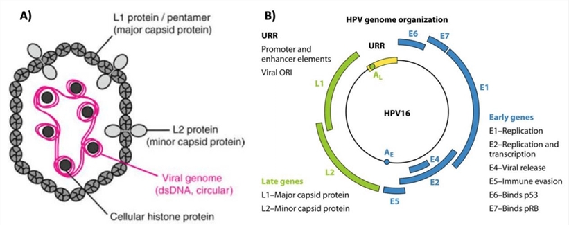
  1. The early (E) region (E1, E2, E3, E4, E5, E6, E7)
  2. The late (L) region (L1 and L2)
  3. The long control region (LCR)

HPV structure and genome organizationFig. 1 HPV structure and genome organization (Modrow, et al. 2010 and Stanley. 2012)

  • E1 uses ATP to exert a helicase activity that forces apart the DNA strands, thus preparing the viral genome for replication by cellular DNA replication factors.
  • E2 facilitates the binding of E1 to the viral origin of replication. E2 also utilizes a cellular protein known as Bromodomain-4 (Brd4) to tether the viral genome to cellular chromosomes.
  • E3 gene exists only in a few papillomavirus types. The gene is not known to be expressed as a protein and does not appear to serve any function.
  • E4 protein of many papillomavirus types is thought to facilitate virion release into the environment by disrupting intermediate filaments of the keratinocyte cytoskeleton. E4 also participate in arresting cells in the G2 phase of the cell cycle.
  • E5 are hydrophobic proteins that destabilize the function of many membrane proteins in the infected cell. The E5 proteins seem to activate the signal cascade initiated by epidermal growth factor upon ligand binding.
  • E6 mediate the degradation of p53, a major tumor suppressor protein, reducing the ability to respond to DNA damage. E6 has also been shown to target other cellular proteins, thereby altering several metabolic pathways.
  • E7 protein is to inactivate members of the pRb family of tumor suppressor proteins. Together with E6, E7 serves to prevent apoptosis and promote cell cycle progression, thus priming the cell for replication of the viral DNA.
  • L1 spontaneously self-assembles into pentameric capsomers. Purified capsomers can go on to form capsids, which are stabilized by disulfide bonds between neighboring L1 molecules. L1 capsids assembled in vitro are the basis of prophylactic vaccines against several HPV types.
  • L2 cooperate with L1 to package the viral DNA into the virion. L2 has been shown to interact with a number of cellular proteins during the infectious entry process.

HPV Vaccine

HPV mainly transmits between individuals through sexual contact and also can spread from a mother to her baby during pregnancy. HPV vaccination provides safe, effective, and lasting protection against the HPV infections that most commonly cause cancer. Serum antibodies against many different viral products have been demonstrated. The best characterized antibodies are those directed against conformational epitopes of the L1 capsid protein assembled as VLPs. There are currently three licensed HPV prophylactic vaccines: Cervarix®, a bivalent HPV-16/18 product from GlaxoSmithKline; Gardasil®, a quadrivalent HPV-6/11/16/18 product and Gardasil®9, a nonavalent HPV-6/11/16/18/31/33/45/52/58 vaccine, both from Merck & Co., Inc. All three vaccines are based on recombinant type-specific L1 capsid proteins assembled into VLPs as immunogens.

References

  1. Sangar VC, Ghongane B, Mathur G. (2016). Development of Human Papillomavirus (HPV) Vaccines: A Review of Literature and Clinical Update. Reviews on Recent Clinical Trials. 11(4), 284-289. 
  2. Deschuyteneer M, Elouahabi A, Plainchamp A, et al. (2010). Molecular and structural characterization of the L1 virus-like particles that are used as vaccine antigens in Cervarix™, the AS04-adjuvanted HPV-16 and -18 cervical cancer vaccine. Human Vaccines. 6(5), 407-419.
  3. Modrow S, Falke D, Truyen U, et al. (2010). Auflage ed. Heidelberg: Spektrum Akademischer Verlag. Molekulare Virologie. 461.
  4. Stanley MA. (2012). Epithelial Cell Responses to Infection with Human Papillomavirus. Clinical Microbiology Reviews. 5(2), 215-220.
  5. Pinto LA, Dillner J, Beddows S, et al. (2018). Immunogenicity of HPV prophylactic vaccines: Serology assays and their use in HPV vaccine evaluation and development. Vaccine. 36(32 Pt A), 4792–4799.
<

Product List

HPV 16 Antigens
Catalog#Product NameExpression System
DAGF-229Recombinant HPV type 16 L1 protein (VLP)E. coliInquiry    
DAG1573Recombinant HPV type 16 [GST]E. coliInquiry    
DAGF-094Recombinant HPV16 E6 protein [His]E. coliInquiry    
DAGF-095Recombinant HPV16 E6 protein (aa 1-158) [His]YeastInquiry    
DAGF-096Recombinant HPV16 E7 protein (aa 1-98) [GST]E. coliInquiry    
HPV 18 Antigens
Catalog#Product NameExpression System
DAGF-230Recombinant HPV type 18 L1 protein (VLP)E. coliInquiry    
DAGF-097Recombinant HPV18 E6 protein (aa 1-158) [His]E. coliInquiry    
DAGF-098Recombinant HPV18 E7 protein (aa 1-105) [His]YeastInquiry    
Inquiry Basket
loading Loading ......

Inquiry

Inquiry