
Inquiry Basket
 Loading ......
Researchers identified Nerve Growth Factor (NGF), which is a critical neurotrophin family member in 1952. NGF's regulation of neuronal survival together with differentiation and regeneration establishes it as a foundational element within neurodevelopmental research. Recent research shows that NGF's effects reach beyond neural functions as it plays an active role in bone metabolism and both inflammatory responses and pain signal transmission. Within bone tissue NGF adjusts the functional equilibrium of osteoblasts and osteoclasts through TrkA and p75NTR receptor activation which results in increased bone formation. It functions as an essential mediator in chronic bone pain development which demonstrates its dual impact on both physiological maintenance and pathological functions.
The polypeptide protein complex NGF occurs widely in human body systems including the brain, ganglia, heart as well as fibroblasts, smooth muscle cells and glial cells. The protein features α, β, and γ subunits while the β subunit containing 118 amino acids functions as the primary center for biological activity. NGF engages with two different receptor types which include the high-affinity tyrosine kinase receptor TrkA and the low-affinity p75 neurotrophin receptor. The TrkA receptor serves as the key receptor responsible for generating beneficial biological effects once it connects with its ligand. This interaction starts intracellular signaling pathways which stimulate the differentiation and development of neurons. TrkA expression occurs throughout all bone-forming cell types which highlights the essential part NGF plays in both skeletal development and homeostasis.
 Figure 1. Neurotrophins and their receptors
(Source: Denk F, et al. 2017)
NGF controls neuronal growth and repair by activating PI3K-AKT and Ras-MAPK signaling pathways. The expression of NGF varies across bone regions with rat ribs showing much higher levels than femurs which might be due to differences in innervation density. NGF promotes faster callus formation during fracture recovery by increasing the expression of bone morphogenetic protein (BMP). NGF triggers osteoblast proliferation and differentiation and facilitates skeletal repair through TrkA receptor-based neurovascular coupling mechanisms.
NGF receptors are widely distributed in bone tissue and its innervating neurovascular structures. Early studies reported that NGF enhances neural innervation and bone regeneration in fracture models, thereby accelerating callus ossification. Through TrkA receptor signaling, NGF directs sensory nerve axons and neovasculature to primary ossification sites, positively regulating osteogenesis via synergistic promotion of vascularization and neural ingrowth. Studies show mature cortical bone produces minimal NGF under normal physiological conditions because NGF is mainly found in the periosteum. Bone damage leads to significant NGF upregulation and prolonged increase within callus tissue which shows its essential role in skeletal development and healing.
Following stress fractures, reinnervation of nerve fibers occurs early within the fracture callus. Most cells in the callus express NGF, guiding the ingrowth of nascent nerves and blood vessels, leading to callus expansion. As overall NGF expression declines, neurovascular ingrowth slows, with residual NGF becoming concentrated in osteoblasts to initiate callus ossification. Pharmacological inhibition of NGF reduces reinnervation, impairs revascularization, suppresses osteoblast activity, and significantly delays callus mineralization.
Bone formation key cells known as osteoblasts show increased alkaline phosphatase activation and type I collagen output when exposed to low NGF levels which promotes cell multiplication. The secretion of NGF from mature osteoblasts during mechanical loading activates osteogenic genes like RUNX2 and BMP2 through TrkA signaling thus enhancing bone mineralization. Research experiments show that when NGF works alongside TGF-β and FGFs they produce faster healing of bone structures. NGF binds to membrane receptors on various target cells to control bone cell growth and differentiation as well as metabolism and apoptosis which leads to changes in skeletal physiology and pathological responses.
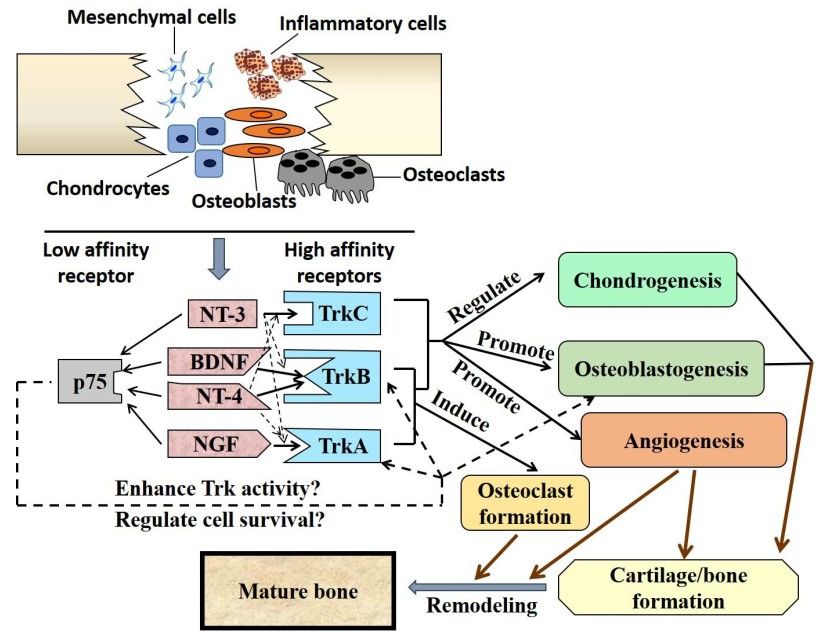 Figure 2. Current knowledge on potential roles of neurotrophins in skeletal injury healing process
(Source: Su YW, et al. 2018)
Proper bone remodeling necessitates equilibrium between bone creation during osteogenesis and bone degradation by osteoclast activity. NGF participates in this regulation through dual mechanisms: NGF activates bone marrow mesenchymal stem cells to become osteoblasts and simultaneously induces osteoclast maturation through pathways that do not involve RANKL. The primary cells that resorb bone tissue originate from monocyte/macrophage hematopoietic lineages and develop into multinucleated giant cells called osteoclasts. Multinucleated osteoclasts display simultaneous expression of both NGF and its receptor TrkA. NGF supports osteoclast development into mature cells which helps regulate excessive bone growth and ensures balanced bone remodeling. NGF produces distinct effects on osteoclast function that depend on dosage levels and environmental conditions including cytokine combinations and mechanical forces.
Skeletal pain results from nociceptive stimuli that activate peripheral pain-sensing neurons and research shows that NGF concentrations rise during tissue damage, inflammatory responses, and persistent pain states. NGF-mediated pain results from a series of events started by the activation of peripheral nociceptors through heat, chemicals or physical stimuli that lead to NGF production and its subsequent release. The NGF-receptor complex enters cells through endocytosis after binding to the TrkA receptor and then moves backwards to neuronal cell bodies where it increases the production of pain-related molecules including bradykinin receptors, voltage-gated sodium channels, and TRPV1. Peripheral sensitization and hyperalgesia result from these receptors while NGF enhances pain signals by activating TRPV1 channels and increasing neuropeptide release including substance P and CGRP to produce peripheral and central sensitization. NGF drives mast cells to release histamine and serotonin along with more NGF which creates a cycle that intensifies pain signaling. In osteoarthritis and similar pathological conditions IL-1β and other inflammatory cytokines trigger NGF overexpression which keeps pain pathways active and leads to continuous sensitization. The complex interaction between NGF/TrkA signaling establishes it as a vital target for treating chronic musculoskeletal pain.
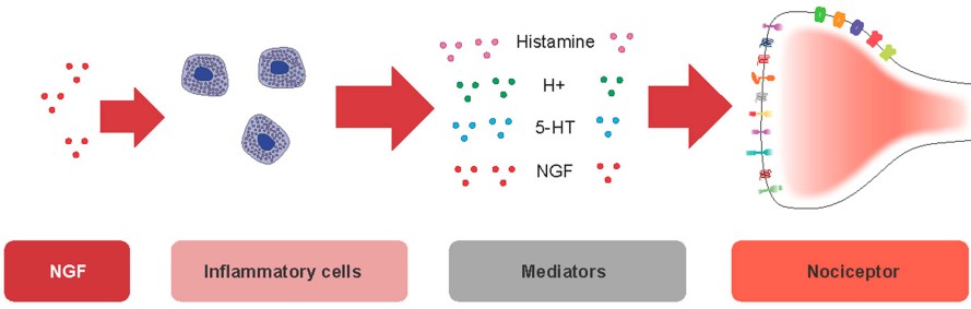 Figure 3. Nociceptive effects of NGF on inflammatory cells
(Source: Barker PA, et al. 2020)
Pain, a hallmark symptom of OA, correlates strongly with elevated NGF expression in affected patients. Research findings demonstrate that increased NGF levels correlate with higher pain severity in knee OA while indicating that NGF overexpression leads to more intense pain and joint function loss. The inflammatory mediator IL-1β advances cartilage breakdown during OA by reducing extracellular matrix components while boosting matrix metalloproteinase (MMP) synthesis. Research findings show that IL-1β provokes dose-related elevation in NGF mRNA and protein levels in chondrocytes. Similarly, IL-1β stimulation of cartilage progenitor cells and synovial tissues markedly elevates NGF production.
NGF interacts with cytokines including TNF-α and TGF-β outside nociceptive pathways. Through the ALK5-Smad2/3 signaling pathway TGF-β triggers NGF expression in human and animal chondrocytes which studies show is more effective than IL-1β for NGF production. The results highlight NGF's functional capacity to serve as both an inflammatory mediator for pain and a cytokine-controlled agent in the development of OA.
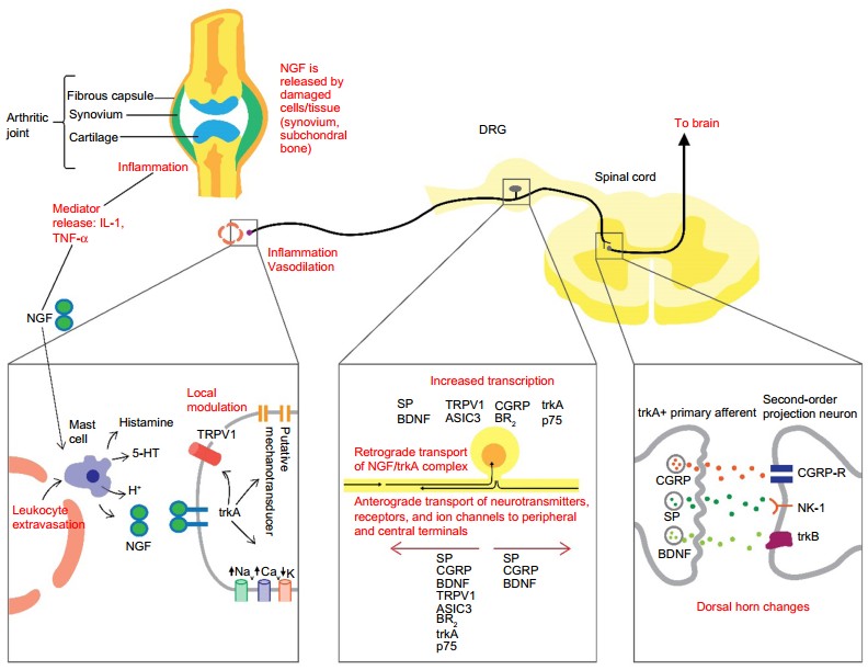 Figure 4. Schematic diagram of the NGF mechanisms involved in the initiation and maintenance of pain
(Source: Chang DS, et al. 2016)
Therapeutic agents specifically developed based on OA pathogenesis help manage pain by neutralizing free NGF molecules and by blocking NGF-receptor binding or preventing TrkA activation. Tanezumab shows strong effectiveness in managing OA pain as part of anti-NGF antibody treatment. Anti-NGF therapy decreases CGRP expression in dorsal root ganglia and improves gait irregularities in animal research while demonstrating its possible benefits and risks for chronic pain treatment. The activation of TRPV1 channels results in increased calcium ion entry and raises the levels of calcitonin gene-related peptide and substance P which leads to central sensitization. TRPV1 agonists show potential as therapeutic agents for managing OA pain.
Pathological Consequences of Dysregulated NGF Levels:
Current challenges lie in balancing NGF's osteogenic benefits against its pain-inducing effects. Combination therapies like NGF inhibitors with NSAIDs or localized sustained-release delivery systems can reduce systemic side effects. Therapeutic outcomes will improve through the development of personalized dosing schedules and biomarker tracking (such as VEGF and BMP levels).
Antibodies that target NGF in Phase II clinical trials block both NGF and TNF-α pathways to improve treatment results and minimize the threat of rapidly progressive osteoarthritis. CRISPR-based gene-editing technologies could enable accurate control of NGF expression leading to innovative treatments for genetic bone diseases. Working together across orthopedics, neuroscience, and pharmacology is essential. Hyperbaric oxygen therapy can advance neural repair through increased NGF expression and 3D biomaterials containing NGF along with mesenchymal stem cells show promise in improving cartilage regeneration. The combination of clinical research with fundamental scientific studies will expedite the development of practical applications in this domain.
References
| Target | Cat. No. | Product Name | Size | Species | Application | Detection Sample | |
| NGF | DEIA7800 | Human NGF/NGFβ(Beta-nerve growth factor) ELISA Kit | 96T | Human | Quantitative | Serum, plasma, tissue homogenates and other biological fluids | Inquiry |
| DEIA753 | Mouse NGF/NGFβ(Nerve growth factor) ELISA Kit | 96T | Mouse | Quantitative | Serum, plasma, tissue homogenates and other biological fluids | Inquiry | |
| DEIA754 | Rat NGF/NGFβ(Beta-nerve growth factor) ELISA Kit | 96T | Rat | Quantitative | Serum, plasma, tissue homogenates and other biological fluids | Inquiry | |
| DEIA135 | Human β-NGF ELISA Development Kit | 15 plates, 45 plates | Human | Quantitative | TBD | Inquiry | |
| ABPR-ZB313 | Human NGF Antibody Pair Set | 5 Plates, 15 Plates | Human | sELISA | Inquiry | ||
| NGFR | DEIA5697 | Human NGFR(p75 neurotrophin receptor) ELISA Kit | 96T | Human | Quantitative | Serum, plasma, tissue homogenates and other biological fluids. | Inquiry |
| Target | Cat. No. | Product Name | Expression System | Tag/Conjugate | Application | |
| NGF | DAG337 | Recombinant Human NGF Protein | CHO Cells | Unconjugated | SDS-PAGE, ELISA | Inquiry |
| DAG-WT3479 | Recombinant Mouse Beta-NGF Protein | CHO cells | N/A | N/A | Inquiry | |
| DAG-WT3480 | Recombinant Human Beta-NGF Protein | HEK293 cells | N/A | N/A | Inquiry | |
| DAG-WT3481 | Recombinant Human Beta-NGF Protein | E. coli | N/A | N/A | Inquiry | |
| NGFR | DAG-P1831 | Rat NGFR peptide | N/A | Unconjugated | ELISA | Inquiry |
| CDBP2036 | Human NGFR blocking peptide | N/A | Unconjugated | Apuri, BL, ELISA | Inquiry |
Loading ......
