
Inquiry Basket
 Loading ......
With the use of secretion inhibitors such as monensin or brefeldin A, secreted cytokines and other proteins can be retained intracellularly. These proteins thus become available for antibody staining, upon fixation and permeabilization of the cells. In general, short-term stimulation of cells with mitogen or antigen is required to induce cellular activation and production of cytokines. One common application of this technique is the visualization of antigen-specific T cells in PBMC or whole blood. This requires stimulation with protein antigens or, commonly, pools of overlapping peptides spanning a protein sequence of interest. The latter, when designed with sufficient length and overlap between peptides, can efficiently stimulate both CD4 and CD8 T-cell responses.
A common protocol for antigen-specific stimulation of T cells for intracellular cytokine staining (ICS) is as follows. Whole blood or PBMC are incubated with antigen or peptide mixtures for 6–16 h. Brefeldin A and/or monensin is added at the time of stimulation (for peptides) or after 2 h (for proteins, to allow for intracellular antigen processing, which is compromised by the secretion inhibitor). At the end of the stimulation period, cells can be held at 4–18°C until ready to process. They are then treated with EDTA to remove adherent cells, fixed (usually with formaldehyde), permeabilized (usually with a detergent), and stained for intracellular determinants. In some cases, surface marker staining is done in conjunction with intracellular staining (this usually works well for CD3, CD4, and CD8). However, most other cell-surface markers require staining prior to fixation, because the epitopes recognized by staining antibodies are sensitive to fixation and/or permeabilization.
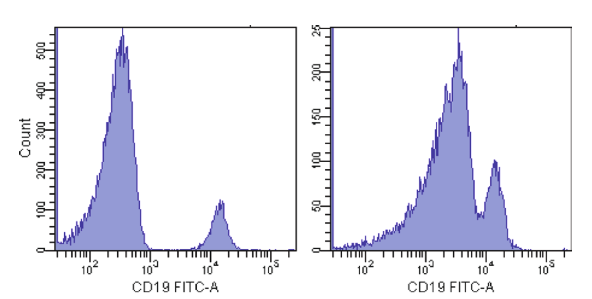 Figure 1. Effect of a bright signal in AmCyan on resolution sensitivity in FITC.
Intracellular staining for multiple cytokines is now often combined with staining for other functional and phenotypic markers as well. This has been made possible by the availability of flow cytometers with digital signal processing, and detectors for up to 18 colors. Along with this instrumentation, software for automated calculation of compensation between colors is now routinely used, often in combination with single-stained capture beads that make construction of compensation controls easier and more precise (since the actual experimental antibodies can be used for compensation, an important consideration for some tandem dye conjugates). Finally, software and fluorescent beads to automate instrument setup and track performance over time are now available, making longitudinal standardization of experiments, at least for a single instrument, much easier. Standardization across instruments, especially given the degree of instrument customization seen in the field, can still be difficult, however.
Despite the advances in tools for multicolor flow cytometry, designing optimal antibody panels of 8 or more colors can be a challenge. The optical spectrum is limited, such that addition of new fluorescent reagents tends to create more spillover into existing detectors. In some cases, this can severely compromise the ability to use those detectors for measurements requiring high-resolution sensitivity.
A general discussion of rules for antibody panel design, along with suggestions for specific fluorochrome combinations and panels, is given in. These rules are very briefly summarized here.
There are also practical considerations to panel design, such as what antibody conjugates are commercially available. In general, it is best to use direct fluorochrome conjugates for multicolor work and for intracellular staining, since nonspecific binding can be a significant concern in these situations.
Even direct antibody conjugates can be optimized by titration for a particular application. Optimal titers should be picked on the basis of maximal signal: noise, which is often obtained below the titer recommended by the manufacturer.
While panels will constantly be refined to include new markers of interest, a degree of standardization is helpful, to avoid extensive re-optimization of new panels and provide for some degree of longitudinal comparisons.
Finally, optimal detection of certain marker combinations requires modification of stimulation and processing steps (e.g. CD107 or CD154).
Reference
Loading ......
