
Inquiry Basket
 Loading ......
A variety of biochemical methods exist for studying protein– protein interactions in mammalian cells, including those based on fluorescence-activated cell sorting (FACS). Strouboulis and colleagues developed a method in which a recombinant protein is tagged with a peptide that is biotinylated in vivo by the coexpressed Escherichia coli BirA biotin ligase. A strength of this approach is the very high affinity interaction between biotinylated substrates and streptavidin (Kd=10−15), which allows high stringencies to be employed during purification. The biotinylation tagging method described here has been successfully used for the single-step purification of a number of transcription factor complexes in nuclear extracts of mammalian cells.
In the initial version of the method, expression cassettes encoding the tagged protein of interest and BirA were subcloned into separate plasmids that also carried a selectable drug resistance gene, the neomycin phosphotransferase gene and the puromycin N-acetyltransferase gene, respectively. Typically, target cells would be transfected with the BirA plasmid by physical methods, such as electroporation, stable clones obtained by the selection of puromycin resistance, and then screened for BirA expression by Northern or Western blot analysis. An appropriate BirA expressing clone would then be transfected with the vector encoding the tagged protein of interest and stable cells expressing the tagged protein biotinylated by BirA obtained by selection for cells that were resistant to the neomycin analog geneticin as well as to puromycin. A drawback of this experimental design is that the optimal concentration of drugs used for the selection depends on the cell line and needs to be determined a priori. Moreover, the selection for drug resistance of the transfected plasmids does not necessarily ensure the coexpression of BirA or the tagged gene of interest.
Hoang and colleagues reported a modification of this methodology in which the tagged gene of interest was coexpressed from a lentiviral vector on a bicistronic transcript that also contained the BirA gene. In addition, the BirA gene was expressed as a fusion protein with the green fluorescent protein (BirA-GFP), allowing stably transduced cells coexpressing the tagged gene of interest and BirA to be isolated by FACS. As described below, we have further modified the FACS-based strategy of Hoang and colleagues. The expression system we have developed consists of two components: a lentiviral vector coexpressing the gene of interest containing a biotinylation tag together with a downstream yellow fluorescent protein (YFP) gene via an encephalomyocarditis virus internal ribosome entry site (IRES); and a second lentiviral vector expressing GFP-BirA.
Our work is focused on TLX1 (T-cell leukemia homeobox 1, previously known as HOX11 or TCL3), an evolutionarily conserved member of the dispersed NKL (NK-Like or NK-Linked) subclass of homeobox genes. The murine ortholog of human TLX1 is essential for splenogenesis and required for the development of certain neurons. Although TLX1 is not expressed in the hematopoietic system, its inappropriate activation is a recurrent event in human T cell acute lymphoblastic leukemia (T-ALL). The manner in which deregulated TLX1expression induces neoplastic conversion remains to be fully elucidated. Several lines of evidence indicate that TLX1 functions as a transcriptional regulator that can either activate or repress gene expression via direct or indirect modes of action. In this regard, TLX1 has been reported to form protein– protein interactions with other transcription factors as well as with a number of transcriptional coregulators and chromatin-modifying enzymes. Among the molecules that have been identified are: CTF1, a ubiquitous transcription factor that associates with TFIIB and the basal transcription machinery; MEIS and PBX members of the TALE (three amino acid loop extension) superclass of homeodomain proteins; the acetyltransferase coactivator CREB-binding protein; the serine/threonine phosphatases PP1 and PP2A; and the eukaryotic initiation factor 4E (eIF4E).
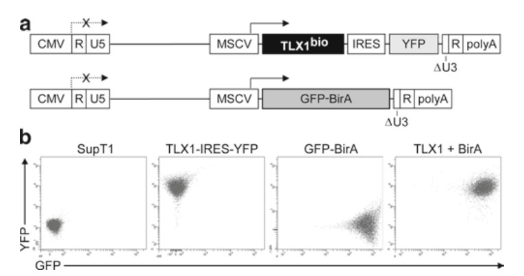 Figure 1. FACS-based binary in vivo biotinylation tagging system.
We recently implemented the in vivo biotinylation tagging approach to isolate and characterize the various TLX1 protein complexes in T-ALL cells. Our initial attempts to coexpress biotinylation-tagged TLX1 and GFP-BirA from a bicistronic lentiviral vector were unsuccessful because the cells did not tolerate high levels of TLX1. Since BirA was expressed on the same transcript, it was selected against. As a result, much higher expression levels of BirA were achieved with the empty GFP-BirA vector, making it difficult to obtain similar levels of endogenous biotinylated proteins for comparisons between the experimental and control samples. To circumvent these problems, we designed a two-component expression system using the simian immunodeficiency virus (SIV) lentiviral vector backbone pCL20cSLFR MSCV-GFP. In pCL20cSLFR MSCV-GFP, which was constructed from the nonpathogenic SIVmac1A11 isolate, the GFP gene is expressed from an internal promoter derived from the long terminal repeat (LTR) of the murine stem cell virus (MSCV), which is highly active in most mammalian cell types. A biotinylation tagging vector expressing a COOH-terminal tagged TLX1 (TLX1bio) under the control of the MSCV LTR was created by inserting the TLX1 coding region in frame upstream of the coding sequences for the BirA target peptide linked to an IRES-YFP cassette, generating pCL20cSLFR MSCV-TLX1bio-IRES-YFP (component 1). A BirA expression vector was similarly constructed by replacing the GFP gene of pCL20cSLFR MSCV-GFP with GFP-BirA, generating pCL20cSLFR MSCV-GFP-BirA (component 2).
In pilot studies, SupT1 cells, which are negative for TLX1 expression but which are arrested at the same stage of T-cell differentiation as TLX1+ T-ALL cells, were transduced with recombinant CL20cSLFR MSCV-TLX1bio-IRES-YFP lentiviral vector particles and/or recombinant CL20cSLFR MSCV-GFP-BirA lentiviral vector particles and stable cell lines obtained by sorting for YFP and/or GFP fluorescence. SupT1 cells expressing GFP-BirA alone served as control for the binding of any biotinylated endogenous proteins to the streptavidin beads. Using single-step affinity capture on streptavidin beads, followed by matrix-assisted laser desorption ionization time-of-flight (MALDI-TOF) mass spectrometry, we identified the Groucho/transducin-like Enhancer of split (Gro/TLE) family member TLE1 as an in vivo binding partner of TLX1. Gro/TLE proteins are regulated by multiple signaling cascades and serve as corepressors for different families of transcription factors. The transcription factors that interact with Gro/TLE corepressors contain short peptide sequences related to either WRPW or to FXIXXIL (where X is any amino acid), the latter referred to as the Engrailed homology 1 (Eh1) motif, a repression domain first identified in the Drosophila Engrailed homeodomain protein. We demonstrated that TLX1 interacts with TLE1 in vitro and in vivo through a seven amino acid sequence encompassing amino acids 19–25 (FGIDQIL) that exhibits similarity to an Eh1 motif. Moreover, we found that this motif was required for optimal regulation of expression of two TLX1 target genes, Aldh1a1 and Fhl1. A previously reported in vitro TLX1 interacting protein, eIF4E, is also efficiently precipitated by streptavidin affinity capture of in vivo TLX1bio protein complexes. Thus, we believe that our adaptation of the FACS-based in vivo biotinylation tagging system provides a powerful tool for the characterization of transcription factor and other protein–protein complexes in mammalian cells.
Transient Transfection
Collection and Concentration of Vector Particles
Titration of Vector Particles
293T (293 human embryonic kidney cells expressing simian virus 40 [SV40] large tumor [T] antigen) cells are highly transfectable such that transient cotransfection with the self-inactivating SIV lentiviral vectors, packaging plasmids and envelope plasmids yields high-titer, replication-defective vector particles. Lentiviral vectors pseudotyped with the VSV-G glycoprotein have a broad host-cell range and can be utilized to transduce all cell types.
Transient Transfection
Collection and Concentration of Vector Particles
Collect the culture medium containing vector particles 24–48 h after medium change. Centrifuge at 2,000×g for 10 min to remove cellular debris and filter through a 0.45 mm pore-size filter (depending on the volume, use a 150-mL filter unit or a small filter unit attached to a 5-mL syringe). Use directly for transductions or aliquot and store at −80°C (see Note 7). Several procedures have been developed to concentrate lentiviral vector particles. The choice of concentration protocol depends on the envelope selected to pseudotype the particle and the quantities of particles to be produced. The stability of the VSV-G envelope protein allows the generation of high-titer lentiviral vector particles by ultracentrifugation, as described here.
Titration of Vector Particles
The HT1080 human fibrosarcoma cell line can be used to determine the titer of the lentiviral vector particles.
The following protocol is used to transduce suspension cell lines with the recombinant biotinylation tagging lentiviral vectors. For adherent cells, the protocol for titration of vector particles is used.
The protocol described below is a variation of the Dignam procedure for the preparation of nuclear extracts for human tissue culture cells that has been modified for T-ALL cell lines (e.g., SupT1). The standard procedure is based on a starting cell number of 2×10⁸ cells and can be adjusted to accommodate 2×10⁹ cells by increasing the buffer volumes at steps 2 and 3 by a factor of 4. All solutions are kept on ice; prior to cell harvesting, freshly thawed PMSF and DTT, and freshly prepared protease and phosphatase inhibitors are added to the prechilled buffers. Centrifugations are performed at 4°C.
Streptavidin affinity precipitation is performed essentially as described (see Note 12).
Reference
Loading ......
