
Inquiry Basket
 Loading ......
Poliovirus (PV) belongs to the family Picornaviridae, Enterovirus group C, and can cause disabling disease. Polio caused by PV is a form of acute flaccid paralysis, and one of the distinguishing features of the infection is that survivors of acute illness may be disabled for life. In rare cases patients die as a result of paralyzed control of the throat or breathing muscles. Since its discovery, PV has been studied extensively to better understand its life cycle, limit transmission and treat polio. PV can spread in cultured non-neural cells, and as a human pathogen, it not only contributed to the invention of the polio vaccine, but also greatly advanced molecular virology as a unique field of research. It was the first animal RNA virus for which the whole genome sequence was determined and for which reverse genetics techniques were developed; the first animal RNA virus for which an infectious clone was constructed and, together with a related rhinovirus, the first human virus for which the three-dimensional structure was solved by X-ray crystallography.
The PV is 25 to 30 mm in diameter, and the viral proteins VP1, VP2, VP3, and VP4 form a capsomer, with 60 capsomers forming the icosahedral viral capsid. The eight protein chains are arranged in β-sheets to form a β-barrel, which constitutes each of the four virions. The different proteins are mixed to form cyclic structures, which act as antigenic sites that bind to the appropriate antibody. There are three serotypes of PV, types 1, 2, and 3. The prototypical strains of type 1 are the Brunhilde and Mahoney strains, the prototypical strains of type 2 are the Lansing and MEFI strains, and the prototypical strains of type 3 are the Leon and Saukett strains.
The PV genome is a single RNA molecule with positive (mRNA-like) polarity and a length of 7.4kb. The viral genome contains a single open reading frame (ORF), 5' and 3' untranslated regions (UTR). Of these, the ORF encodes a polyprotein of about 2200 residues, which is ultimately converted into 11 mature polypeptides by hydrolytic activity, with some intermediate stages of processing acting as discrete functional units. The last four polypeptides (VP1-VP4) correspond to the N-terminus of the polypeptide and are structural components of the viral capsid. The remaining polypeptides are involved in viral genome replication, hydrolytic processing of polypeptide proteins, and a variety of activities that directly or indirectly ensure the efficient generation of viral progeny. In addition, viral RNAs contain a number of cis-acting components, the replication elements oriL, oriR, and oriI in the 5UTR and 3UTR, which hijack the cellular translation machinery. The translational cis-element in the 5UTR, called the internal ribosomal entry site (IRES), is responsible for cap-independent internal initiation of translation of the viral RNA.
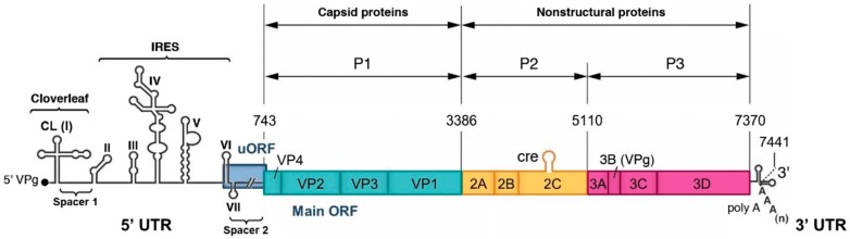 Figure 1. Organization of the PV1 genome
(Source: Quarleri J. 2023)
The variability of the PV genome is a key feature underpinning the evolution of the virus and its pathogenicity and epidemiological profile. It is also an important tool for understanding the link between genotype and phenotype. Covalent modifications of viral RNA are caused by both replicative and non-replicative processes. The 3D gene encodes a viral RNA-dependent RNA polymerase (RdRP) that replicates viral RNA with the help of other viral and host proteins. The enzyme may have problems with premature termination and incorporation of incorrect nucleotides, in addition to point mutations, intra- or intermolecular rearrangements, and other replication errors that can lead to mutations. When using purified enzyme preparations to measure the extent of nucleotide misincorporation, the error frequency values were in the range of 5×10⁻³- 10-5, with transitions occurring approximately ten times more frequently than transversions. Each progeny RNA molecule produced by RdRP carries, on average, a nucleotide variant different from its template, but the degree of infidelity of poliovirus RdRP fluctuates. PVs with more accurate RdRP decayed more rapidly, were less competitive, and were less adaptable.
In 1949 researchers discovered that PV could multiply in cultured non-neural human cells, and later isolated PV genomic RNA was shown to be infectious to monolayers of HeLa cells, suggesting that the viral genome itself is a biologically active vector that leads to infection. PVs utilize RdRP for replication, and their genome is translated into a very large polypeptide that is later specifically cleaved into smaller functional proteins. Meanwhile, cellular fractionation studies have revealed that poliovirus RNA is synthesized in replication complexes that bind different membrane structures in the cytoplasm of infected cells. In 1977 VPg was discovered, a small protein consisting of 22 amino acids covalently linked to the 5' end of poliovirus RNA. The absence of VPg when the viral genome is translated on a multimer suggests that VPg is removed before or during translation, and its presence at the 5' end of newly synthesized positive- and negative-stranded RNA suggests that VPg may be involved in the initiation of RNA synthesis.
IRES allows the host translation apparatus to recognize viral mRNAs in the absence of a 5' cap structure on the viral mRNA, which could allow PVs to compete effectively with the cellular translation machinery through a cap-independent mechanism. The cis-replication element (Cre) in the PV genome is an RNA stem-loop structure located almost entirely within the coding region and is required for viral RNA replication. These elements bind to viral proteins involved in the formation of RNA replication complexes, thereby specifically recognizing viral RNA in the cytoplasm of infected cells among the myriad of host cell mRNAs containing poly(A). Cre sequences promote uridylation of VPg, the primer that initiates viral RNA synthesis, via 3Dpol. Their function appears to be strand-specific, as Cre is required for positive-strand RNA synthesis, whereas negative-strand RNA synthesis may not require Cre.
For PV, a class of viruses with very limited coding capacity, the ability to usurp cellular components and structures for their own benefit during infection is important. Lysis of components of the nuclear pore complex (e.g., Nup153 and p62) by the viral 2A protease results in the accumulation of a number of proteins normally found in the nucleus in the cytoplasm of virus-infected cells. These relocalized nuclear proteins typically have RNA-binding capabilities and play a role in the RNA metabolic steps of the host cell. For example, the cellular and protein SRp20 was relocated to the cytoplasm of PV-infected human cells, and it is an important IRES trans-acting factor in PV translation. TDP2 acts as a DNA repair enzyme that breaks the protein-RNA covalent linkage of VPg at the 5' end of viral RNA. It is thought to be the source of VPg unchaining enzyme activity.
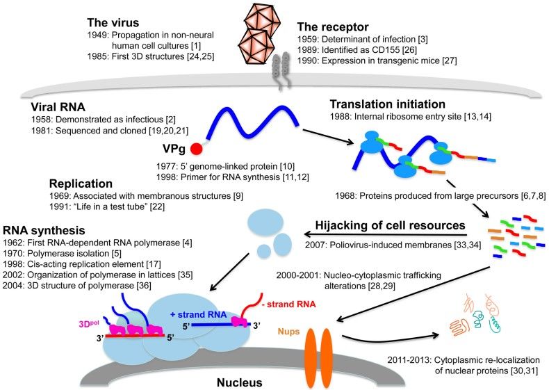 Figure 2. Important findings on the PV replication cycle over the last 65 years
(Source: Lévêque N, et al. 2015)
The only known natural host of PV is humans, and it is generally spread by the fecal-oral route. After localized growth in the tonsils and cervical lymph nodes, the virus spreads to Peyer's patches and the small intestine. The virus multiplies in the pharynx and intestine for 1 to 3 weeks, with a typical incubation period of 2 to 35 days. The local immune response usually prevents the spread of the virus, so most patients with the infection are either asymptomatic or characterized by flu-like symptoms. Gastroenteritis, respiratory infections, and flu-like illnesses may have self-limiting episodes. Antibodies may cause a decrease in viremia and may also be transferred to the central nervous system (CNS) through the bloodstream. The most virus is excreted in the feces 2 to 3 days before and 1 week after the onset of symptoms. PV has a specific affinity for the cellular receptor CD155 that propagates along afferent nerves in the brain. In humans, CD155 protein has been detected in intestinal epithelial cells, M cells in Peyer plaques, and germinal centers within Peyer plaques.
 Figure 3. Pathogenesis of poliomyelitis
(Source: Mbani CJ, et al. 2023)
The anterior horn cells of the spinal cord are severely damaged by the cytopathic effect of PV, which leads to paralysis of the patient's limbs. Posterior horn cells of the spinal cord, thalamic motor neurons, and hypothalamus are potential targets for virus transmission. Brainstem involvement can be fatal. The histology of damaged brain cells showed vacuolization and infiltration with aggregates of plasma cells, polymorphonuclear neutrophils and microglia. Macrophages phagocytose infected cells, leading to axonal degeneration. Muscle atrophy spreads widely, leading to flaccid paralysis. Post-polio syndrome (PPS) may develop 25 to 30 years after the first episode of paralysis. Persistent degeneration of motor neurons in patients with PPS may be responsible for the progressive muscular dystrophy symptoms seen in patients with PPS.
Two vaccines are currently used to prevent polio caused by PV, the injectable inactivated polio vaccine (IPV) and the oral polio vaccine (OPV).
OPV, also known as Sabin vaccine, initially contained three live attenuated strains of polio, and a PV strain with reduced neurotoxicity was developed by utilizing an attenuation technique whereby the virus is continuously passed on in non-human primates and cultured primate cells. Vaccination with OPV is effective in preventing fecal shedding of wild-type PV after exposure, and it prevents PV infection by inducing sufficient intestinal mucosal immunity. OPV aims to eradicate polio by preventing the transmission of the virus from person to person. However, a long-standing disadvantage of using OPV is that attenuated vaccine strains may revert to a neuropathogenic phenotype during replication in the gut. In areas with low vaccination coverage, these pathogenic OPV-derived strains may have the ability to spread among local populations, maintaining a reservoir of pathogenic strains and causing polio.
IPV, administered by intramuscular injection, does not induce sufficient intestinal immunity to effectively prevent transmission of the virus. IPV induced higher and near 100% seroconversion rates for all three poliovirus serotypes after three doses of vaccination, and IPV reduced the duration and rate of viral shedding in immunized individuals compared with unimmunized individuals. Vaccination with IPV significantly improves mucosal immunity in OPV-vaccinated children, and the effect may be greater than that of additional OPV vaccination. Despite the excellent safety profile of IPV, silent transmission of wild-type PV may occur in countries where only IPV is used.
References
| Target | Cat. No. | Product Name | Size | Species | Application | Detection Sample | |
| Poliomyelitis Virus | DEIA372 | Human Anti-Polio Virus 1-3 IgG ELISA Kit | 96T | Human | Quantitative | Serum, Plasma or other Biological Fluids | Inquiry |
| DEIASL265 | Simian Anti-Poliovirus Type 1 IgG ELISA Kit | 96T | Quantitative | Srerum | Inquiry | ||
| DEIASL266 | Simian Anti-Poliovirus Type 2 IgG ELISA Kit | 96T | Quantitative | Srerum | Inquiry | ||
| DEIASL267 | Simian Anti-Poliovirus Type 3 IgG ELISA Kit | 96T | Quantitative | Srerum | Inquiry | ||
| PVRL1 | DEIA-FN1231 | Human PVRL1 (Poliovirus receptor-related protein 1) ELISA Kit | 96T | Quantitative | Serum, plasma, cell culture supernatants, tissue homogenate | Inquiry | |
| DEIA-FN1232 | Mouse PVRL1 ( Poliovirus receptor-related protein 1) ELISA Kit | 96T | Quantitative | Serum, plasma, cell culture supernatants, tissue homogenate | Inquiry |
Loading ......
