
Inquiry Basket
 Loading ......
Influenza virus is called influenza virus for short. The typical clinical symptoms of these epidemic diseases are acute high fever, body pain, significant fatigue and respiratory symptoms. Influenza virus is mainly transmitted through droplets in the air, contact between susceptible and infected persons or contact with contaminated objects. Generally, autumn and winter are its peak seasons. Human influenza is mainly caused by influenza A virus and influenza B virus. Influenza virus has weak resistance to the outside world. Animal influenza virus usually does not infect humans, and human influenza virus usually does not infect animals, but pigs are an exception. Pigs can be infected with both human influenza virus and avian influenza virus, but they are mainly infected with swine influenza virus. A few animal influenza viruses can cause human influenza pandemics after adapting to humans.
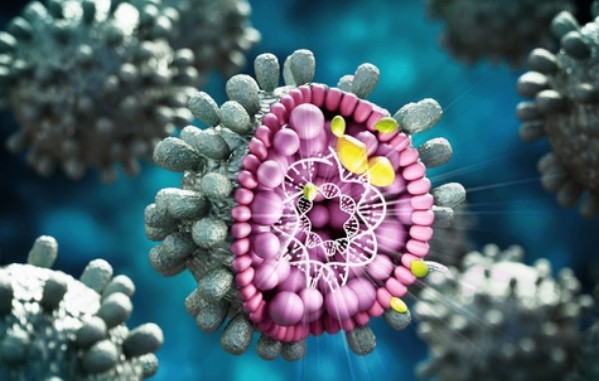
The influenza virus possesses a segmented genome and a helical capsid structure while existing as single-stranded negative-sense RNA. human influenza viruses can be divided into three categories according to the antigenicity of their nucleoproteins:

Influenza A virus
Influenza B virus

Influenza C virus
The respective roles of these proteins include facilitating viral entry into host cells and enabling viral release from the same cells. Influenza B viruses are classified into two lineages: The Influenza B virus consists of two lineages B/Yamagata and B/Victoria yet Influenza C appears less frequently and produces milder symptoms.The immune system targets the HA protein as its main focus and this protein drives both antigenic drift and shift of the virus.
The structure of influenza virus can be divided into three parts from the outside to the inside: envelope, matrix protein and core.

The core of the virus contains the genetic material that stores the virus information and the enzymes necessary to replicate this information. The genetic material of influenza virus is single-stranded negative-strand RNA, abbreviated as ss-RNA. ss-RNA is combined with nucleoprotein (NP) and entangled into ribonucleoprotein body (RNP), which exists in a very high density. In addition to ribonucleoprotein body, there is also RNA polymerase responsible for RNA transcription.

The matrix protein constitutes the outer shell skeleton of the virus. In fact, in addition to the matrix protein (M1), there is also membrane protein (M2) in the skeleton. M2 protein has the function of ion channel and regulating the pH value in the membrane, but the number is very small. The matrix protein is bound to the outermost envelope of the virus to protect the virus core and maintain the spatial structure of the virus.

The envelope is a layer of phospholipid bilayer membrane wrapped outside the matrix protein. This membrane comes from the host's cell membrane. The mature influenza virus buds from the host cell, wraps the host's cell membrane around itself, and then detaches from the cell to infect the next target. In addition to phospholipid molecules, there are two very important glycoproteins in the envelope: hemagglutinin and neuraminidase.
| Transmission and Pathogenesis | Descriptions |
| Source of Infection | The main source of infection is patients, followed by latently infected persons, and animals carrying the virus may also be the source of infection. |
| Transmission Route | Droplets carrying influenza viruses enter the body through the respiratory tract, and a few people can also be infected through indirect contact such as sharing handkerchiefs and towels. |
| Infectivity | After the virus enters the population, it is highly contagious and can spread rapidly. The speed and breadth of transmission are related to population density. If the virus that enters the human body is not cleared by the cough reflex, or is not neutralized by the body's specific IgA antibodies and inactivated by nonspecific inhibitors in mucosal secretions, it can infect a small number of respiratory epithelial cells, causing the cells to produce vacuoles and degenerate. |
| Impact | The virus's NA can reduce the viscosity of the respiratory mucus layer, not only exposing the cell surface receptors, which is conducive to the adsorption of the virus, but also promoting the spread of virus-containing fluids to the lower respiratory tract, causing many respiratory cells to be damaged in a short period of time. Influenza viruses generally only cause surface infections, but not viremia. |
Influenza diagnosis depends on both medical examination and laboratory examinations. During the clinical assessment doctors evaluate patient symptoms together with information about their recent flu exposure and influenza outbreak status in the community. Viral infection can also induce the expression of interferon and cellular immune regulation, causing some autoimmune reactions, including high fever, headache, gastrocnemius and systemic muscle pain, etc. The toxin-like products of viral metabolism and the products released by cell necrosis can also cause and aggravate the above reactions. It is not difficult to diagnose influenza in combination with clinical symptoms during the epidemic period, but laboratory tests must be performed for diagnosis or epidemic monitoring, mainly including virus isolation and culture, serological diagnosis and rapid diagnosis methods.
| Method | Descriptions |
| Isolation and identification of viruses | Usually, a hemagglutination test is performed on the throat washing fluid or throat swab of patients within 3 days of onset. If the hemagglutination test is positive, a hemoagglutination inhibition (HI) test is performed with a known immune serum to identify the type. If the hemagglutination test is negative, the chicken embryo is used for blind passage for 3 more times. If hemagglutination still does not occur, the virus isolation is judged to be negative. Tissue culture cells (such as human embryonic kidney or monkey kidney) can also be used to isolate the virus. T |
| Serological diagnosis | Double serum samples are taken from patients in the acute phase (within 5 days of onset) and the recovery phase (2 to 4 weeks of disease course), and the HI test is often used to detect antibodies. If the antibody titer of the recovery phase serum is more than 4 times higher than that of the acute phase, a diagnosis can be made. Normal human serum often contains non-specific inhibitors, so the serum can be treated with trypsin before the HI test to avoid affecting the HI test results. |
| Rapid diagnosis | Rapid diagnosis of patients mainly uses indirect or direct immunofluorescence and ELISA to detect viral antigens. Patients' nasal mucosal prints or respiratory epithelial cell smears are often taken, and immunofluorescence staining with fluorescein-labeled influenza virus immune serum is used to check antigens, or ELISA is used to check antigens in patients' throat gargles. |
To prevent and control influenza virus, on the one hand, we need to strengthen the detection of influenza virus mutations and make accurate predictions as much as possible so as to carry out targeted vaccination; on the other hand, we need to cut off the spread of influenza virus in the population. Influenza virus is transmitted by droplets. Early detection of influenza patients and fumigation of public places with chemical disinfectants can effectively inhibit the spread of influenza virus. For influenza patients, interferon, adamantane, oseltamivir and other drugs can be used for treatment. Interferon is a cytokine that can inhibit viral replication. Adamantane can act on influenza virus membrane protein and hemagglutinin protein to prevent the virus from entering host cells. Oseltamivir can inhibit neuraminidase activity to prevent mature viruses from leaving host cells.
Research indicates that immunization with an HA construct without the globular head domain triggers a wider immune response while protecting against various influenza subtypes. Protection from lethal influenza virus challenges resulted when mice received vaccinations featuring a headless HA construct according to animal model research.The development of vaccines targeting the M2 protein represents another promising strategy since this protein plays a role in viral replication processes. Studies demonstrate that focusing on the M2 protein enables the creation of vaccines with broad protective capabilities. Research demonstrates that virus- like particles (VLPs) effectively generate protective immunity for both identical and different strains of the influenza virus. Virus-like particles (VLPs) reproduce the virus structure yet lack replication ability and stimulate potent immune responses without posing any disease risk.
The high mutation rate of the influenza virus presents substantial public health difficulties because it can lead to pandemic outbreaks. Today's vaccines and antiviral treatments offer essential protection but face limitations because of the virus's changing nature. Creating a universal influenza vaccine represents an essential objective for advancing influenza research and public health. The use of conserved viral regions combined with advancements in mRNA and VLP technologies enables scientists to create vaccines that can protect against various influenza subtypes.
| Cat. No. | Product Name | Host | Isotype | Application | |
| DPAB-L21083 | Anti-IAV H1N1 Neuraminidase Polyclonal antibody | Rabbit | IgG | I-ELISA | Inquiry |
| DPAB-L21217 | Anti-IAV H1N1 Neuraminidase Polyclonal antibody | Rabbit | IgG | I-ELISA | Inquiry |
| CABT-B8414 | Anti-Influenza A H9N2 (A/Hong Kong/1073/99) Neuraminidase polyclonal antibody | Rabbit | IgG | ELISA | Inquiry |
| CABT-BL1383 | Anti-EPN1 (aa 431-480) polyclonal antibody | Rabbit | IgG | WB, ELISA | Inquiry |
| CABT-BL8953 | Anti-Parainfluenza Virus Type 3 p69 polyclonal antibody | Goat | / | IFA | Inquiry |
| DPAB-CS24038A | Human Anti-PIV IgA Control Serum | Human | IgA | ELISA | Inquiry |
| CABT-CS679 | Rabbit Anti-IAV H5N8 HA Polyclonal Antibody | Rabbit | IgG | WB, ELISA | Inquiry |
| Cat. No. | Product Name | Size | Species Reactivity | Application | Detection Method | |
| DEIA-CL036 | Influenza A Virus Nucleoprotein Antigen ELISA Kit | 96T | IAV | Quantitation | sELISA | Inquiry |
| DEIA353 | Influenza A IgG ELISA Kit | 96T | Human | Quantitative and Qualitative | ELISA | Inquiry |
| DEIA358 | Influenza B IgM ELISA Kit | 96T | Human | Quantitative and Qualitative | ELISA | Inquiry |
| Cat. No. | Product Name | Size | Target | Species | |
| DAG-P2864 | PIV type 1 Nucleocapsid (aa 0-0) | / | / | PIV | Inquiry |
| DAG-P2865 | PIV type 3 Nucleocapsid (aa 0-0) | / | / | PIV | Inquiry |
Loading ......
