Introduction of Indirect ELISA Protocol
First described by Engvall and Perlmann, the enzyme-linked immunosorbent assay (ELISA) is a technique based on an enzymelabeled antibody capable of detecting and quantifying protein immobilized to a solid surface. ELISA is traditionally performed in 96-well or 384-well polystyrene plates where antigen or antibody binds through passive absorption. As the technology advances, variations to ELISA such as ELISPOT and in-cell ELISA emerge. Multiplex microarray ELISA significantly improves the traditional technique by allowing each well to screen multiple analytes with a limit of detection 1,000-fold higher than traditional ELISA. The term ELISA now loosely applies to assays involving antibody detection of analyte.
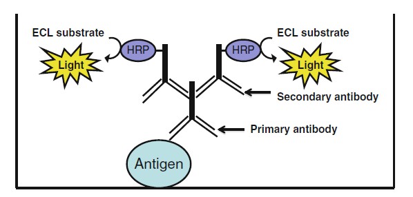 Figure .1 Schematic diagram to illustrate the principle of indirect ELISA.
Traditional ELISA can be performed in four different formats, direct, indirect, sandwich, and competitive. In indirect ELISA, the antigen of interest is immobilized to a 96-well or 384-well polystyrene microtiter plates by passive absorption. A blocking buffer is added to saturate all unbound sites in the well followed by incubation with an unlabeled primary antibody specific for the antigen. An anti-species enzyme-conjugated secondary antibody is subsequently added to bind to the primary antibody. Typically, the secondary antibody is conjugated to horseradish peroxidase (HRP) and detected with enhanced chemiluminescent substrate (ECL). Another common method is to utilize biotin-coupled primary antibody and enzyme-conjugated streptavidin secondary antibody. In both cases, the substrate produces measurable unit corresponding directly to the amount of antigen present in the well. This chapter describes the methodology of performing indirect ELISA on 96-well polystyrene plates using HRP-conjugated secondary antibody followed by detection with enhanced chemiluminescent substrate (ECL). In addition, procedures to optimize the antigen, primary antibody, and secondary antibody by chessboard titration are also provided. Similar to direct ELISA, indirect ELISA is useful for antibody screening, epitope mapping, and protein quantification. The secondary antibody serves to enhance the signal of the primary antibody thus making it more sensitive than direct ELISA. However, it also produces a higher background signal and potentially decreases the overall net signal. As with any immunoassay, optimization is required.
Methods of Indirect ELISA Protocol
Antigen Immobilization
- Dilute antigen in binding solution. Antigen should be diluted into a final concentration ranging from 1–100 μg/ml.
- Add 100μl of diluted antigen into each well. Seal the plate with cover or adhesive and incubate for 2 h at room temperature or overnight at 4 °C (see Note 3).
- Aspirate off the antigen and wash four times with 200 μl wash solution either by hand or ELISA plate washer. This step washes off any unbound antigen.
- Optional step: short chemical denaturation (see Note 4).
(a) Prepare 1 M solution of guanidine-HCl.
(b) Add 100 μl to each well and incubate for 1 h.
(c) Aspirate off the antigen and wash four times with 200 μl of wash solution either by hand or ELISA plate washer.
Blocking
- Add 200 μl of blocking solution into each well and incubate at room temperature for 30 min to overnight (see Note 5).
- Aspirate off the blocking solution. It is not necessary to wash the plate at this point.
Incubation with Primary Antibody
- Dilute primary antibody in dilution buffer. Primary antibody should be diluted into a final concentration of 1μg/ml or follow manufacturer’s instruction.
- Add 100μl of diluted HRP-conjugated antibody into each well. Seal the plate with cover or adhesive and incubate at room temperature for 1 h.
- Aspirate off the antibody and wash four times with 200μl wash solution either by hand or ELISA plate washer.
Immobilized Antigen Detection by HRP- Conjugated Secondary Antibody
- Dilute HRP-conjugated secondary antibody in dilution buffer. HRP-conjugated antibody can be commercially purchased and is usually anti-species to the primary antibody. The secondary antibody should be diluted into a final concentration following manufacturer’s instruction or 1:10 K of 1 mg/ml stock.
- Add 100 μl of diluted HRP-conjugated antibody into each well. Seal the plate with cover or adhesive and incubate at room temperature for 1 h.
- Aspirate off the antibody and wash four times with 200μl wash solution either by hand or ELISA plate washer.
- Prepare ECL substrate according to manufacturer’s instruction.
- Add 50–100 μl of substrate into each well.
- Place the ELISA plate on a titer plate shaker and incubate for 2 min at room temperature.
- Read the light signal (relative light units) using PerkinElmer VICTOR™ X3 Multilabel Plate Reader.
Optimization by Chessboard Titration
It is essential to optimize each component of indirect ELISA to avoid prozone or hook effect and generate reliable data. Chessboard titration is a common method allowing the titration of two reagents at a time. This section describes the steps for optimization of antigen, primary antibody, and HRP conjugated secondary antibody concentrations.
Chessboard Titration of Antigen and Primary Antibody
Titration of Antigen
- Fill columns 2–12 with 100μl of binding solution.
- Dilute antigen in binding solution into a final concentration of 100 μg/ml for partially purified protein sample or 10μg/ml for purified small peptides.
- Add 200μl of diluted antigen sample into all eight wells of column 1.
- Aspirate off 100μl of diluted antigen from wells in column 1 and transfer to wells in column 2. Gently pipette up and down three times to mix the content of the well.
- Repeat step 4 and perform dilute antigen from columns 2–11. Leave column 12 with binding buffer only.
- Seal the plate with cover or adhesive and incubate for 2 h at room temperature or overnight at 4 °C.
- Aspirate off the antigen and wash four times with 200μl wash solution either by hand or ELISA plate washer.
- Add 200μl of blocking solution into each well and incubate at room temperature for 30 min to overnight.
- Aspirate off the blocking solution. It is not necessary to wash the plate at this point.
Titration of the Primary Antibody
- Fill rows B–H with 100μl of dilution buffer.
- Dilute the primary antibody in dilution buffer into a final concentration of 1 μg/ml.
- Add 200μl of diluted antibody into all 12 wells of row A.
- Aspirate off 100μl of diluted antibody from row A and transfer to row B. Gently pipette up and down three times to mix the content of the well.
- Repeat step 4 and titrate primary antibody from row B to row G. Leave row H with dilution buffer only.
- Seal the plate with cover or adhesive and incubate for 1 h at room temperature.
- Aspirate off the antigen and wash four times with 200 μl wash solution either by hand or ELISA plate washer.
Detection with HRP- Conjugated Secondary Antibody
- Dilute HRP-conjugated secondary antibody in dilution buffer. The secondary antibody should be diluted into a final concentration following manufacturer’s instruction or 1:10 K of 1 mg/ml stock.
- Add 100μl of diluted HRP-conjugated antibody into each well. Seal the plate with cover or adhesive and incubate at room temperature for 1 h.
- Aspirate off the antibody and wash four times with 200μl wash solution either by hand or ELISA plate washer.
- Prepare ECL substrate according to manufacturer’s instruction.
- Add 50–100μl of substrate into each well.
- Place the ELISA plate on a titer plate shaker and incubate for 2 min at room temperature.
- Read the light signal (relative light units, RLU) using PerkinElmer VICTOR™ X3 Multilabel Plate Reader.
Chessboard Titration of Primary Antibody and HRPConjugated Secondary Antibody
Titration of Primary Antibody
- Dilute antigen in binding solution. Antigen should be diluted into the optimal concentration obtained from chessboard titration of antigen and primary antibody. 12.5 μg/ml for partially purified protein sample and 1.25 μg/ml for purified peptides.
- Add 100 μl of diluted antigen into all 96 wells. Seal the plate with cover or adhesive and incubate for 2 h at room temperature or overnight at 4 °C.
- Aspirate off the antigen and wash four times with 200 μl wash solution either by hand or ELISA plate washer. This step washes off any unbound antigen.
- Add 200 μl of blocking solution into each well and incubate at room temperature for 30 min to overnight.
- Aspirate off the blocking solution. It is not necessary to wash the plate at this point.
- Fill columns 2–11 with 100 μl of dilution buffer.
- Dilute primary antibody in dilution buffer into a final concentration of 1 μg/ml.
- Add 200 μl of diluted primary antibody into all eight wells of column 1.
- Aspirate off 100 μl of diluted primary antibody from wells in column 1 and transfer to wells in column 2. Gently pipette up and down three times to mix the content of the well.
- Repeat step 7 and dilute primary antibody from columns 2–11. Leave column 12 with dilution buffer only.
- Seal the plate with cover or adhesive and incubate for 1 h at room temperature.
- Aspirate off the antigen and wash four times with 200 μl wash solution either by hand or ELISA plate washer. This step washes off any unbound primary antibody.
Titration of HRP Conjugated Secondary Antibody
- Fill rows B–H with 100 μl of dilution buffer.
- Dilute the HRP-conjugated antibody in dilution buffer into a final concentration fourfold higher than recommended by the manufacturer or 1:2,500 of 1 mg/ml stock solution.
- Add 200 μl of diluted antibody into all 12 wells of row A.
- Aspirate off 100 μl of diluted secondary antibody from row A and transfer to row B. Gently pipette up and down three times to mix the content of the well.
- Repeat step 4 and titrate secondary antibody from row B to row G. Leave row H with dilution buffer only.
- Seal the plate with cover or adhesive and incubate for 1 h at room temperature.
- Aspirate off the antigen and wash four times with 200 μl wash solution either by hand or ELISA plate washer.
- Prepare ECL substrate according to manufacturer’s instruction.
- Add 50–100μl of substrate into each well.
- Place the ELISA plate on a titer plate shaker and incubate for 2 min at room temperature.
- Read the light signal (relative light units) using PerkinElmer VICTOR™ X3 Multilabel Plate Reader.

 Loading ......
