
Inquiry Basket
 Loading ......
Immunohistochemistry (IHC) is a critical ancillary test that aids in the accurate diagnosis of the central nervous system neoplastic and nonneoplastic conditions, this method localize and demonstrate specific antigens of interest in tissues or cells based on antigen antibody interactions visualizing it with the aid of a chromogen and light microscopy.
The use of IHC has been a routine and indispensable facet of modern diagnostic cancer pathology practice. Newer IHC biomarkers are being continuously found as indispensable aids in differential pathologic diagnosis, prognosis, or prediction of response to molecular-targeted therapies.
The workflow of IHC begins with the preanalytic phase (i.e., cold ischemia) followed by analytic phase (e.g., antigen retrieval) and postanalytic phase (microscopic interpretation), with a strict quality control to reach optimal results.
This chapter focuses on the materials, methods, and techniques used in immunohistochemistry and the principles of quality control and validation.
Fixation and Cold Ischemia
The most widely used fixative is 10% neutral buffered formalin (NBF). This fixative is an aldehyde fixative that is categorized as a "cross-linking" fixative. This fixative has a good preservation of morphology, of antigens and for small molecules such as hormones.
The time of fixation (6–72 h), the volume (10 times the specimen) and the cold ischemic time (less than 1 h) are critical for an optimal staining and to avoid false-positive or false-negative results. See Notes 1 and 2.
Microtomy and Slide Preparation
Tissue sections should be cut at 3–4 μm using charged/PLUS slides. This contributes to good adhesion of the tissue to the slide and accurate postanalytic interpretation. Thicker sections, folds and tears can cause false-positive or false-negative staining results.
Dewaxing and Hydrating
For automated processes it is not strictly necessary any of the changes in xylene and ethanol for dewaxing. Only follow step 1 and insert the section slides into the automated stainer and run a specific protocol.
Epitope Retrieval
Epitope retrieval is referred to unmasking the epitope. Options for epitope unmasking include heat-induced epitope retrieval (HIER) and proteolytic-induced epitope retrieval (PIER)/enzymatic retrieval. PIER was the first method developed to address the antigen-masking effects of formalin fixation, the enzymes used in this technique are protease, proteinase K, trypsin. These methods sometimes produce false-positive or false negative results.
HIER methods have greatly improved the quality and reproducibility of IHC staining on the application of heat to formalinfixed paraffin-embedded (FFEP) section, using retrieval solution of varying pH levels in order to break cross-linkages and expose epitopes. The pH is critical and the heat sources include microwave oven, pressure cooker and water bath or automated IHC staining instrument. Regardless of the method used, do not let the slides dry out.
We use the HIER method with a pressure cooker for manual process with the following steps:
Blocking Nonspecific Binding
Background staining is a frequent problem in IHC and is a cause of nonspecific binding related to endogenous biotin, endogenous enzyme activity, and hydrophobic interactions. If the nonspecific binding is not blocked it could lead to false-positive results. Endogenous avidin binding has been observed in all biotin-based staining methods. Endogenous biotin is bound to enzymes and protein is frequently seen in brain tissue. Blocking is achieved by applying unlabeled avidin followed by an application of unlabeled biotin. If enzymes similar to that of the antibody label are present in the tissue sample being stained, they may react with the substrate and cause background staining.
Hydrophobic interactions result from cross-linking of amino acids and proteins during formalin fixation. Tissues that exhibit background staining due to hydrophobic interactions include collagen, other connective tissues, epithelium, and adipose tissue.
Blocking Method
Antibodies
Some antibodies need to be diluted, the optimal dilution is when the concentration of the antibody produces an adequate staining with minimal background staining and minimal nonspecific binding. The antibodies are diluted with proteinaceous buffered solution known as a diluent.
Other antibodies are "ready to use" (RTU). The benefits of RTU antibodies are their relative lot-to-lot consistency, reproducibility, and exclusion of a possible source of laboratory error during antibody titration.
On the following tables we exemplify some of the antibodies used for glial tumors and medulloblastoma as well as what immunoreaction we expect to find on each tumor.
Method
Secondary Antibody/Detection System
The secondary antibody is used in the indirect method, which is a multistep method with specificity against the unlabeled primary antibody to amplify and visualize the antibody-antigen reaction. This amplification is possible because multiple secondary antibodies bind to a single primary antibody. The sensitivity of detection of target antigens increases with the addition of secondary antibodies.
The direct method is a one-step staining method, in which the primary antibody is conjugated directly to the label. This method lacks sensitivity and that is why it is not widely used.
Method
Chromogen and Substrates
The detection of antigens using an antibody requires a chromogen to visualize the target antigen. The incubation of slides with a chromogen substrate (color-producing system) results in a stable, colored end-product suitable for visualization with light microscopy. Staining in tissue sections results from the reaction of a soluble substrate with an enzyme to produce an insoluble, colored product. The intensity of the color produced when the substrate is added correlates with the concentration of the primary antibody and respective tissue antigen.
The most common enzymes are horseradish peroxidase (HRP) and alkaline phosphatase (AP). The associated chromogens for HRP are 3,30-diaminobenzidene tetrahydrochloride (DAB) and 3-amino-9-ethylcarbazole (AEC).
Method
Counterstaining
The counterstaining is a process that stains the section that did not participate in the histochemical reaction. It is critical to use a counterstain that is compatible with the color produced by chromogen. The most frequent stain used is hematoxylin as a counterstain for a histochemical reaction producing a brown or red color.
Method
Controls
There are two specific types of controls that must be accounted for in each IHC run: positive controls to confirm the analytic sensitivity of the assay (that the assay detected the antigen) and negative "tissue" controls to confirm the analytic specificity of the assay (that the assay is negative in the absence of antigen).
Positive control tissue must contain the target antigen and should be fixed, processed, and embedded in the same manner as patient samples being tested. Also should reflect the intended use of the antibody in diagnostic practice. It is preferable to use a tissue with low levels of antigen staining to confirm adequate analytic sensitivity of the assay, as loss of a significant degree analytic sensitivity may not be detected using tissue with intense immunoreactivity, thus leading to false-negative interpretations of patient tissue samples. It is optimal to have the control sections mounted on each slide of patients' tissue to confirm that the IHC assay worked on that particular slide. See Note 9.
Negative tissue controls are those tissues known to be negative for the antigen of interest and processed in the same manner as test samples are used. Negative tissue controls assess the specificity of the primary antibody by verifying the absence of cross-reactivity. One control block may contain positive and negative tissue for the assay and it is possible to use in the same slide.
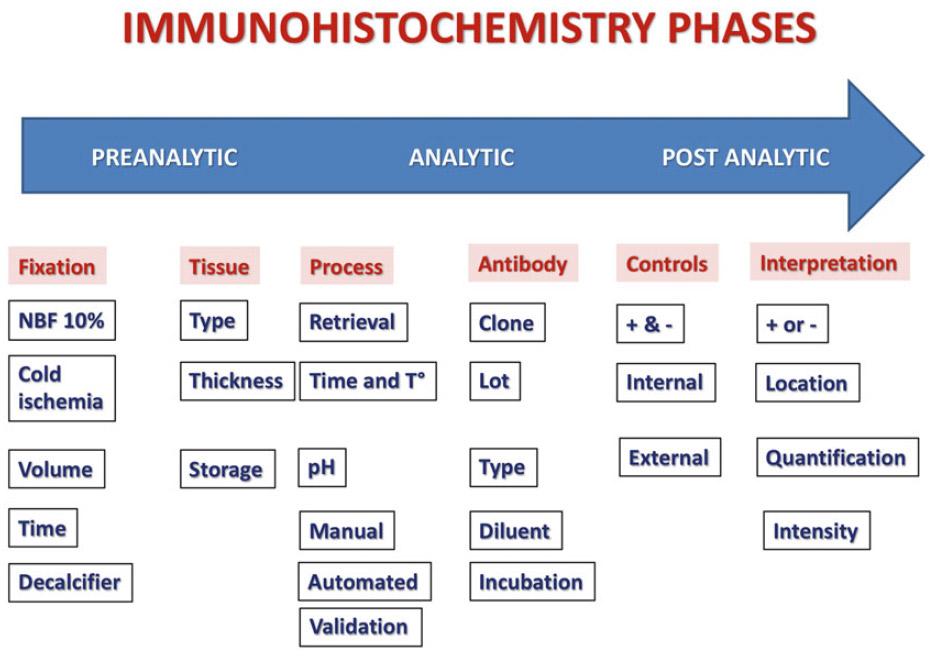 Figure 1. Schematic representation of the steps in the different phases of immunohistochemistry for the Central Nervous System.
Interpretation
The interpretation of the staining depends on the antibody used; it is very important to define the cell localization, the intensity, and sometimes the percentage of positive tumor cells.
The staining in normal tissue elements, if present within the specimen, should be considered as an additional check on the IHC assay performance.
Quality Management
Quality management involves quality control (QC) and quality assurance (QA) programs, as well as quality improvement (QI) initiatives.
QC consists in optimizing each aspect of the IHC, from specimen arrival to reporting the results, and includes the validation of the IHC assay.
QA programs establish the policies and procedures for daily QC and external proficiency testing, in addition to monitoring other quality metrics during preanalytic, analytic, and postanalytic phases.
QI is the practice of continuously assessing and adjusting performance using statistically and scientifically accepted procedures.
Principles of Analytic Validation
The goal of the analytic validation is to define the reproducibility, reliability, and accuracy of the antibodies to detect the molecule or antigen in the tissue before placing it into clinical service.
The basic principles or recommendations for analytic validation are:
Reference
Loading ......
