
Inquiry Basket
 Loading ......
The interferon-gamma (IFN-γ) release assay (IGRA) is an in vitro extension of the century-old in vivo tuberculin skin test, better known as the TST. The TST was one of the first diagnostics and is still used today. The need for a diagnostic replacement for the TST had been long recognized, but awaited scientific advancement. Shortcomings include limitations in specificity and sensitivity. Reduced specificity correlates with the use of the Bacille Calmette-Guérin (BCG), a vaccine given in most countries. TST sensitivity is reduced in persons with immunosuppression, a characteristic that has become more pronounced with the worldwide HIV epidemic.
IGRAs employ scientific achievements in assay development and cytokine detection using either the ELISA or ELISPOT platform to capture IFN-γ produced in response to antigen stimulation within 16–24 h. The first IGRA prototypes, like the TST, relied on cell-mediated responses to purified protein derivative (PPD), a purified fraction of Mycobacterium tuberculosis (MTB) proteins. Sequencing of the MTB genome revealed a region of difference (RD-1) that was not present in BCG. This finding led to the replacement of the IGRA PPD peptide pool with two specific RD-1 MTB peptides (ESAT-6 and CFP10) and recently TB 7.7. The use of RD-1 peptides increased predictive values of IGRAs and further increased their viability as diagnostics. IGRAs also offer improved diagnostic feasibility compared to the TST because there is no requirement for a follow-up visit reducing the cost for the provider, and barriers to care, such as transportation, scheduling, and cost for the subject.
The ELISPOT platform requires isolation of peripheral blood mononuclear cells (PBMCs) from whole blood, a step that requires more time and training. The isolation of PBMCs, however, is essential to both the specificity and sensitivity of the assay and serves a dual purpose. First, it ensures that the same number of PBMCs are stimulated, allowing the assay to be less dependent on the subjects' T-cell number or underlying T-cell repertoire. Secondly, isolation of PBMCs allows spot visualization of individual effector cells. Spot size and density can be used to define the measured kinetics of the secreting cell. This function may prove to be valuable as the specificity of spot readers improves or as scientific knowledge concerning TB immunology advances. Most effector memory T cells secrete their products within 4–6 h of antigen presentation. In the ELISA assay, the secreted IFN-γ may be diluted, degraded, or utilized in proportion to the timing of the antigen-presenting cell (APC) presentation and the stop point in the assay. The ELISPOT immediately captures IFN-γ produced directly from each cell, resulting in the generation of a cellular "footprint" and the reduction of dilution. For a T cell, this footprint has a characteristic dark center with a halo effect that is the visual result of the degradation of the initial IFN-γ produced upon activation. The dimensions and intensity of the generated footprint are indicators of the avidity of the secreting cell.
ELISA-based QuantiFERON-TB Gold® (QFT-G) and QuantiFERON-TB Gold in Tube® (QFT-GIT) and the ELISPOT-based T-SPOT.TB® have gained endorsements from medical entities, such as the American Thoracic Society (ATS) and Center for Disease Control and Prevention (CDC), and approval as a latent TB infection (LTBI) diagnostic from regulatory agencies in many countries, including the US FDA, European CE Mark, and Chinese FDA. Since approval of the first TB IGRA in 2001, and the first M. tuberculosis-specific IGRA in 2004, extensive research has focused on the positive and negative predictive values of each platform. This type of scrutiny is not common for a diagnostic, but is expected because of the global burden of LTBI and the lack of a gold-standard for comparison. Without a gold-standard, those interested in reporting assay performance must explain the meaning of discordance with the TST and the discordance of serial assays. Overall, the use of IFN-γ production as a diagnostic indicator has proven to be useful; but mechanisms that cause variability are imperative for understanding IGRA results in the context of disease progression and coinfection.
Comparatively little research has focused on the cellular mechanisms involved in IGRA variability. The measurement of IFN-γ responses to TB-specific antigens is dependent not only on the platform, but more importantly on the frequency of IFN-γ - producing memory cells, the time since exposure, interleukin-2 (IL-2) and IL-12 availability, effective antigen presentation, which can vary depending on the APC type in vivo, and whether the T cell has been recently activated. We reasoned that since IL-7 is a cytokine known to cause immune activation, affect dendritic cell maturation, and enhance antigen-specific responses and T-cell survival, it might improve memory T-cell responses in vitro.
We stimulated isolated PBMCs in both IGRA platforms, and measured antigen-specific responses with and without IL-7 in IGRA-positive subjects (ESTAT-6 and CFP10 peptides). We also measured responses in both control and IGRA-positive subjects to tetanus, viral peptide pools from influenza virus, cytomegalovirus, Epstein-Barr virus (CEF) and cytomegalovirus (IE-1), negative control peptides, and a positive-control mitogen, phytohemagglutinin (PHA). In both diagnostic platforms, IL-7 augments memory-specific responses. In vitro, IL-7 increases the number of cells producing IFN-γ as measured by the TSPOT.TB ® assay. In ELISA, the cell-to-volume ratio of 250,000 cells in 100 Pl increased the difference between IL-7-treated and -untreated samples. In addition to more spots, some IL-7-augmented responses have an increased production of IFN-γ per cell or "spotforming unit" as measured by spot size comparisons to antigen stimulation without IL-7. The spots produced by IFN-γ-producing cells vary in size. Clonal T-cell pools activated by a single type of APC demonstrate that although the spot sizes of a T cell vary, they follow a lognormal distribution. Software on the automated CTL ELISPOT reader (S4) allows the comparison of histograms of the footprints in each well or in groups of wells. The kinetics of the IFN-γ-secreting cell detected by the ELISPOT membrane is measured by the rate at which IFN-J is released (spot density) and the length of time that the cell secretes (spot size). Histogram comparisons of the category boundaries (the log of the spot size in square millimeters) measured by the spot reader show that the addition of IL-7 increased the length of time that the cell secreted, indicating that APC contact time was increased. There are two likely cellular sources of IFN-γ production in the IGRA assays: NK cells and T cells. Because IL-7 increased the number of spots, it was important to test whether the spots resulted from secreting NK cells. We, therefore, depleted PBMC samples so that the only remaining cell type was NK cells. We also depleted PBMC samples to study isolated T-cell subsets. We found that the footprints made by NK cells were much smaller than those made by T cells. The use of a plate reader (such as CTL) allows for spot gating based on size which can exclude NK cells when measuring T cell-specific responses.
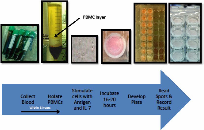 Figure 1. Diagram of process.
Loading ......
