
Inquiry Basket
 Loading ......
Hybridoma technology provides a useful means of generating monoclonal antibodies against any antigenic molecule. The production or isolation of a high-quality immunogen is the first step in the process. This can be accomplished by any number of methods that include: (1) chemical synthesis, (2) purification, (3) peptide synthesis, or (4) recombinant protein expression. An immunogen can be pure or crude and immunogenicity of the target will invariably influence the antigenic response following immunization. Consequently immunogenicity is determined empirically and is often facilitated by integration of large carrier molecules like keyhole limpet hemocyanin (KLH) or bovine serum albumin (BSA), adjuvant, animal, route, and schedule of immunizations. Provided that the immunogen elicits the desired immune response, the strategy used to identify or “screen” hybridomas for the desired monoclonal antibody (mAb) is essential for successful identification and isolation. Toward this aim it is necessary to defi ne what type of mAb is to be identified and the appropriate hybridoma screening method devised to achieve the result.
First consider the myeloma or hybrid cell line to be used as fusion partner. These cell lines are all derived from a single parent Balb/c mouse lineage, yet by subcloning or fusion, they have evolved with different properties that influence cell fusion efficiency and production of extraneous antibodies. We prefer lines that do not produce any immunoglobulins such as the P3X63-Ag8.653 and choose to immunize young postweaning (4–6 weeks) Balb/c mice to maintain isogenic compatibility to minimize potential rejection of cloned hybridoma cell lines used for in vivo production of ascites.
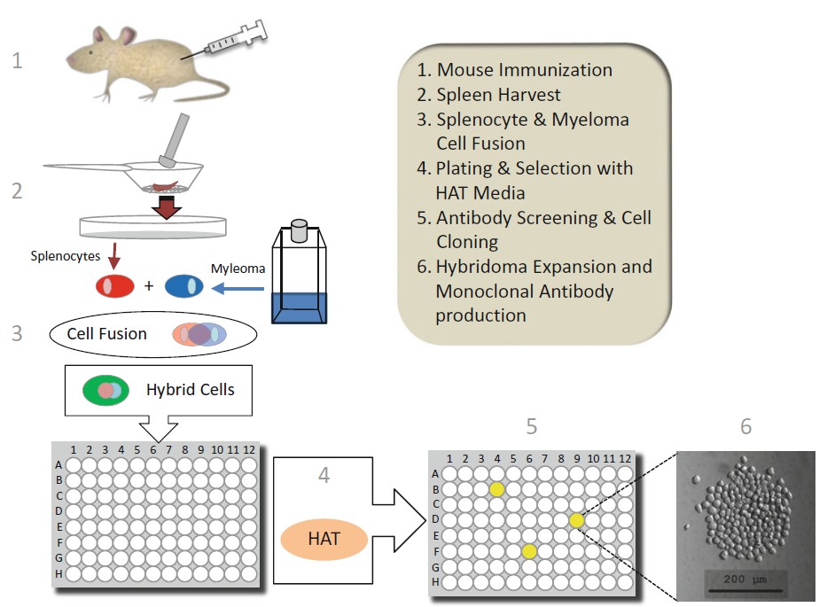 Figure 1. Schematic of hybridoma technology illustrating steps involved in the generation of a monoclonal antibody producing hybridoma cell line.
The immunogen will often defi ne how the hybridoma screen will be performed. With purified or synthetic immunogens, efforts should be made to engineer the best tools to facilitate hybridoma screening for the target. For example, if one is to use the same synthetic peptide as the immunogen and the screening antigen, it is beneficial to use different carrier conjugates for immunization and screening to avoid isolation of antibodies against the carrier. In this scenario you might generate (1) a synthetic 20-mer peptide conjugated to KLH to serve as immunogen and (2) the same 20-mer peptide conjugated to BSA to serve as antigen for screening mAbs (see Note 1). Here you immunize with peptide- KLH and screen mAbs for binding to peptide-BSA immobilized on microtiter plates by indirect ELISA. An alternate scenario is that you immunize with a 20-mer peptide-KLH conjugate and screen against an immobilized crude extract containing the parent protein by iELISA. Similar strategies can be engineered for small chemical molecules or recombinant proteins expressed via DNA expression vectors containing terminal protein tags such as GST or SUMO. The basic principle is that if the immunogen is not “native” the antigen used in the hybridoma screen should differ to avoid isolating mAbs against undesired structures.
One of the simplest designs for isolating antigen uses the indirect ELISA (iELISA) where the target antigen is immobilized on plastic microtiter plates, hybridoma supernatants added to allow any mAb present to bind target, and an anti-mouse antibody conjugated to a reporter used to identify mAb binding to antigen. The commercial availability of isotype-specific secondary antibodies should be considered to avoid isolation of sticky nonspecific or low-affinity IgM antibodies (see Note 2). This approach for the identification of hybridomas producing target mAb is effective, but often the resulting antibody isolated fails to “capture” antigen in solution, an important requirement for at least one of the two antibodies used in the construction of a sandwich ELISA (sELISA). These “detector” types of antibodies are common and are often excellent at binding target antigen in a variety of immunoassay formats such as Western blot, immunohistochemistry, iELISA, or direct ELISA (dELISA). Equally they are usually suitable for use in sELISA as the detector mAb that is directly conjugated to a reporter.
Most high-affinity in-solution capture antibodies are conformational, binding to discontinuous epitopes on the molecule. We believe that the immobilization of a protein antigen can deform the protein and skew the selection of mAbs toward those that best bind linear or continuous epitopes. To isolate a capture mAb usually requires a different hybridoma screening strategy. This usually requires the preexistence of a suitable “detector” antibody to create the pair (see Note 3). Often with novel antigens, these mAbs are not available and one might need to generate a detector mAb first before seeking a capture mAb. Other strategies can be devised that take advantage of recombinant DNA engineering or synthetic peptide chemistry. Here the integration of a protein tag (GST or SUMO) or biotin can be used with a corresponding tag-specific antibody to facilitate identification of a capture mAb.
One example of a basic screen for capture capable mAb for a protein might involve: (1) immobilizing an isotype-specific anti- mouse mAb on microtiter plates, (2) capturing mAb from hybridoma supernatants, (3) adding protein-tagged antigen in solution, and (4) adding non-murine polyclonal secondary detector antibody- conjugated reporter. Several alternatives can be devised that might include the use of an avidin reporter in place of a secondary antibody or immobilized avidin microtiter plates to capture and orient a biotinylated antigen followed by binding of hybridoma supernatant mAb and detection with an anti-mouse conjugated reporter.
It is important to have a hybridoma screening strategy defined to maximize the successful isolation of a mAb that binds target antigen and best meets the end-user need. Often a multitude of hybridomas will be cloned producing individual mAbs with unique antigen-binding properties that perform in some immunoassays and not others. Part of the characterization of an isolated mAb involves evaluation of cross-reactivity, species specificity, affinity, and immunoassay performance.
The basic workflow of generating a mAb using hybridoma technology includes: (1) immunogen design, production, and immunization, (2) cell fusion, (3) hybridoma selection with HAT, (4) hybridoma mAb screening, (5) cell cloning and expansion, and (6) ascite production. After mice are immunized, those with the highest serum antibody titer to the antigen are euthanized and their spleen harvested (see Note 4). The final immunogen boost should be administered into the intraperitoneal cavity without adjuvant 3 days before the cell fusion (see Note 5).
Myeloma cells in culture should be split into fresh media 1 day before the fusion event to ensure log growth and maximal health (see Note 6). Myeloma cells are mixed with splenocytes in the presence of polyethylene glycol (PEG) to change membrane permeability and allow cell fusion. The cell fusion events are random and the culture will contain a mixture of single cells along with splenocyte- splenocyte, myeloma-myeloma, and hybrid myelomasplenocyte.
Myeloma cells lack the enzyme hypoxanthine-guanine phosphoribosyltransferase (HGPRT) which prevents the cells from using the salvage pathway to synthesize purines. Myeloma cells grown in the presence of aminopterin, a folic acid analogue that blocks de novo cell biosynthesis of purines and pyrimidines necessary for DNA synthesis by inhibiting dihydrofolate reductase, will die even with the addition of salvage pathway precursors hypoxanthine and thymidine (HT) given their HGPRT status. Splenocytes express HGPRT and thymidine kinase (TK) enzymes and can utilize the salvage pathway for survival in the presence of aminopterin if supplied with hypoxanthine and thymidine (HT). Primary cells however are not immortal and will die off in time when grown in cell culture. Consequently hybrid cells that result from a fused myeloma-splenocyte retain the myelomacontributed immortality and express HGPRT obtained from the splenocyte allowing only the hybrid cells to survive and grow in HAT (hypoxanthine-aminopterin-thymidine)-supplemented medium.
The purpose of HAT medium is to (a) selectively kill unfused myeloma cells that would otherwise overgrow any fused hybrids generated and (b) eliminate any myeloma-myeloma hybrids that lack HGPRTase. HGPRT-positive splenocytes or splenocytesplenocyte hybrids are not sensitive to HAT by means of salvage pathway support but as a primary cell have a finite lifespan and will die in culture over a short period of time. After HAT selection is complete (10–14 days), cell culture supernatants are screened for mAb, and those cells selected for expansion and cloning are passaged in medium containing hypoxanthine-thymidine (HT) without aminopterin before final expansion in normal hybridoma growth medium.
Laminar flow hood for sterile cell culture.
Autoclave.
Liquid aspirator.
Single and multichannel pipettes.
Clinical swinging bucket centrifuge.
37 °C water bath.
CO2 incubator.
Hot plate.
Hemocytometer.
Timer.
Inverted phase contrast microscope.
Immunized mice that have been boosted with immunogen 3 days prior to the day of cell fusion.
Myeloma cell line, subcultured (split) the day before cell fusion to achieve 2–3 × 10⁵ cells/mL.
Sterile stainless steel cell dissociation sieve with 100 mesh wire screen and pestle.
Sterile 5-mL disposable syringe.
Sterile cell scraper.
100-μm sterile cell strainer (BD Falcon).
500 mL Erlenmeyer flask.
200-mL glass beaker; filled with pre-warmed 37 °C water.
Red blood cell lysis buffer.
Hybridoma grade DMSO.
Sterile 100-mL solution basins.
Clear, sterile, 96-well, cell culture-treated, flat-bottom plates with lids.
Sterile surgical tools that include scissors and forceps.
70 % alcohol.
Sterile plastic petri dishes.
Sterile Pasteur pipettes.
5-, 10-, and 25-mL plastic disposable serological pipettes.
Sterile disposable polypropylene 15- and 50-mL conical tubes with screw top lid.
Solid polyethylene glycol (3,000–3,700 PEG; Sigma—melted, neutralized, and pre-warmed to 37 °C).
1 N HCL.
pH test paper.
Disposable sterile 0.22-μm syringe filter.
Iscove’s medium with L -glutamine and sodium bicarbonate (3.024 g/L).
Iscove’s medium with 10 % FBS.
HAT-selective medium; Iscove’s with 10 % FBS and 1× HAT from 100× stock supplemented with 10 % macrophage-conditioned medium (MCM) ( see Note 7 ).
HT-selective medium; Iscove’s with 10 % FBS and 1× HT from 50× stock.
We prefer to use dry-powered Iscove’s versus premade liquid medium as it has better storage and shelf life. This dry formulation contains L -glutamine but requires the addition of 3.024 g/L of sodium bicarbonate. Medium is dissolved in ultrapure water and bicarbonate added (shift in yellow solution to red). From this stock Iscove’s solution, three medium formulations are made: (1) Iscove’s with 10 % fetal bovine serum (FBS), (2) Iscove’s with 10 % FBS and 10 % macrophage-conditioned media (MCM) and 1 × HAT from 100 × HAT stock, and (3) Iscove’s with 10 % FBS and 1 × HT from 50 × HT stock. All solutions are sterile fi ltered into sterile media bottles (see Note 8).
Place a 5-g bottle of PEG on a hot plate to liquefy the wax. One must monitor this so as not to overheat the PEG. Place a needle through the membrane as a pressure release device so the top won’t pop off. Use “high heat” setting and monitor the melt which should yield ~4 mL. Once liquefied, add 5 mL of stock Iscove’s (no serum) and then add 700 μl of DMSO followed by 1–2 drops of 1 N HCL until slightly acidic/neutral pH is achieved— test using pH strip paper. Filter sterilize the ~10 mL PEG through a 0.22-μm syringe filter into a sterile tube and place in 37 °C water bath until needed.
Immunized mice are anesthetized then euthanized by cervical dislocation and placed in a laminar hood, body saturated in 70 % alcohol, and spleens aseptically removed (trim clean) and placed in a tube containing 25 mL of sterile pre-warmed 37 °C Iscove’s medium with 10 % FBS. Perform one fusion at a time at a steady pace. Open a sterile 100 mm Petrie dish and place the sterile stainless steel sieve with mesh on the bottom of the dish. Pour the spleen and medium into the sieve then using a sterile glass pestle or sterile plunger from a 5 mL syringe mash the spleen through the metal mesh of the sieve toward the bottom of the Petri dish to obtain a cell suspension. Using a sterile pipette, pass and mash fresh media through ~10 mL through the sieve to dislodge and push through cells; leave any fatty connective tissue on the mesh screen. Remove sieve and transfer the dispersed cells in media from the bottom of the dish through a 100 μm sterile nylon cell strainer into a fresh 50 mL sterile conical tube. Add fresh media slowly to the cell strainer and gently mash bits with a sterile 5 mL syringe plunger through the nylon mesh to pass and transfer cell suspension into tube. Centrifuge cells at 1,500 rpm for 5 min in a swinging bucket clinical centrifuge to pellet, aspirate and discard supernatant then gently suspend cell pellet in 10 mL of pre-warmed red cell lysis buffer and incubate for 10 min at 37 °C; at this time begin preparation of myeloma cells. After 10 min incubation in RBC lysis buffer centrifuge splenocytes to pellet and resuspend in Iscove’s medium without FBS. Count cells to determine yield (we routinely recover ~ 10 8 splenocytes).
Myeloma cells grow in suspension forming cell clusters. We use several large flasks to maintain cells for harvest and 1 day before fusion cells are subcultured into fresh media to maximize growth and health. To harvest myeloma cells from culture fl asks we gently scrape the fl ask bottom and transfer the cell suspension into a 50 mL conical tube, centrifuge at 1,500 rpm for 5 min in clinical centrifuge to pellet, aspirate the media and suspend the cells in prewarmed Iscove’s basal medium without FBS and repeat centrifugation. Finally we suspend the cells in fresh Iscove’s without FBS and count cells to determine yield (see Note 9).
Loading ......
