
Inquiry Basket
 Loading ......
The Herpesviridae family consists of enveloped DNA viruses which have more than 200 species that affect mammals and birds as well as bivalves and amphibians. The viruses share identical structural elements which include linear double-stranded DNA inside an icosahedral capsid surrounded by tegument proteins and a lipid bilayer envelope with glycoproteins. The human population has nine herpesviruses that cause infections including HSV-1 and HSV-2, VZV, EBV, CMV, HHV-6A and HHV-6B, HHV-7, and HHV-8. Herpesviruses are further grouped into three subfamilies based on differences in their patterns of infection: Alphaherpesvirinae, Betaherpesvirinae and Gammaherpesvirinae. Alphaherpesviruses have a short replication cycle, replicate rapidly, can infect a wide range of cell types and usually become latent in sensory ganglia. HSV-1, HSV-2 and VZV all belong to the Alphaherpesvirinae subfamily.
Genital herpes represents one of the most widespread sexually transmitted infections which remains persistent and highly contagious because of HSV-2. The virus primarily spreads through sexual contact and maternal transmission results in permanent and untreatable infection. The majority of HSV-2 infections remain without noticeable symptoms although the virus produces recurrent genital ulcers in some cases. The virus continues to shed from genital mucosa even when no visible lesions exist which enables its transmission to other people. Moreover, HSV-2 infection recruits CD4+ T cells to the genital area—cells that serve as targets for HIV—facilitating HIV entry through breaks in the skin. As a result, individuals with HSV-2 infection also face an increased risk of acquiring HIV.
HSV-1 and HSV-2 have linear double-stranded DNA genomes about 100–150 kb in length, surrounded by an icosahedral capsid made up of 162 capsid protein subunits. The complete virus is up to approximately 200 nanometers in diameter, depending on the size of the envelope. The envelope itself contains 13 different glycoproteins, of which gD, gB, and the gH/gL mediate host cell entry. Between the capsid and the envelope is a dense protein layer called the tegument. This contains several proteins involved in viral replication and assembly. HSV-1 and HSV-2 have about 74% homology in nucleotide sequence. The two viral envelopes have different protein compositions that provide antigenic differences.
HSV-1 and HSV-2 are capable of entering a latent stage in peripheral neurons. In the latent state, the viral genome persists but there is no production of virus until reactivation. Primary infection in the natural host is through mucosal surfaces or broken skin. The virus replicates in cells at the site of initial infection. The virus enters the peripheral nervous system through nerve endings in the mucosa or infected skin. The virus is transported by retrograde axonal transport and the viral genome is released into the neuronal nucleus. This process is essential for establishing latency in sensory ganglia and remains inactive until the virus reactivates. Upon reactivation, newly formed viral particles travel anterograde along the nerves back to the mucocutaneous sites they innervate (the epidermis or other nerve-innervated tissues), causing recurrent infection.
 Figure 1. Establishment of HSV latency and reactivation in neurons
(Source: Bai L, et al. 2024)
HSV-1 and HSV-2 show their highest level of productivity through reactivation in the trigeminal ganglia (TG) and the lumbosacral dorsal root ganglia (DRG) ganglia. The herpes simplex virus exists as two subtypes HSV-1 and HSV-2 which differ only slightly in their tissue preferences and antigenic features. HSV-1 is often transmitted through oral–oral contact and is the causative agent of orolabial herpes. HSV-2 is often transmitted by sexual contact and is the causative agent of genital herpes.
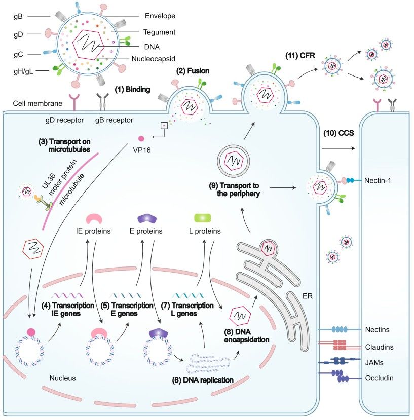 Figure 2. Host cell entry and transmission of HSV
(Source: Bai L, et al. 2024)
HSV-2 lifecycle is composed of five stages: attachment, entry, capsid nuclear import, genome replication and assembly, and release. Attachment is mediated by viral glycoproteins on the envelope and their interaction with host-cell surface Heparan sulfate proteoglycans (HSPGs). Binding to HSPGs is mediated by gB and gC. Once gB and gC tether HSV-2, gD binds to a series of entry receptors including HVEM, nectin-1/2, and 3-O-HS and gH/gL and gB together mediate fusion between viral envelope and the cell membrane or receptor-mediated endocytosis. The nucleocapsid becomes free in the cytoplasm after this process. The capsid moves through the cytoskeleton network of microtubules and motor proteins until it reaches a nuclear pore where Hsp90 and other host factors help release linear double-stranded DNA into the nucleus. The viral genome enters the nucleus where it quickly forms a circular shape before starting its gene expression sequence of immediate-early (IE) followed by early (E) and late (L) genes. The IE genes receive regulation from VP16 and host RNA polymerase II while E gene products including thymidine kinase start viral DNA replication and L genes produce structural proteins. The virus forms new capsids inside the nucleus before it exits through the inner nuclear membrane and sheds its envelope when it reaches the cytoplasm. The virus obtains its final envelope by entering Golgi-derived vesicles during the budding process which results in the formation of mature virions.
HSV-2 infection exists in two forms—lytic infection and latent infection—which dynamically alternate. The primary infection of HSV-2 begins by infecting epithelial cells before it travels to nerve endings and moves retrograde through axons to establish latency in the sacral ganglia. The virus moves anterograde from the nerve cells to skin and mucosal surfaces to produce genital ulcers during reactivation. The two infection states exist through chromatin modifications which enable lytic infection through H3K9ac/H3K14ac and H3K4me2/3 histone modifications for immediate-early gene expression but latent infection through LAT-induced heterochromatin marks (H3K9me2/3, H3K27me3) that suppress lytic gene expression.
The immune system can reach its full potential against HSV-2 through administered vaccines which activate both humoral and cellular immunity to eliminate HSV from the body. Scientists use mice and rabbits as animal models to study HSV infection responses and pathogenic mechanisms and vaccine efficacy because mice help analyze acute infection and latency and neuronal infection while rabbits help study viral reactivation. The complex genomic structure of HSV-2 together with its infection process and potential for latent reactivation creates challenges for developing HSV-2 vaccines because animal models fail to perfectly replicate human infection. The research goals determine if HSV-2 vaccines belong to therapeutic or prophylactic categories because therapeutic vaccines target HSV-2–infected individuals to reduce symptoms and infectivity while prophylactic vaccines protect HSV-2–naive populations from infection. The HSV-2 vaccine candidates fall into four main categories which include inactivated vaccines and subunit vaccines and replication-defective vaccines and live-attenuated vaccines and DNA vaccines and peptide vaccines.
The HSV vaccine based on recombinant protein subunits utilizes gB and gD as its immunogenic components. The two glycoproteins generate neutralizing antibodies and they produce immune responses that protect against both HSV-1 and HSV-2 infections. The gD2/gB2 recombinant subunit vaccine which uses HSV-2 glycoproteins stands as the leading candidate among all available options. The identification of antigens which detect HSV-infected cells while preserving cell health remains difficult and researchers need to find new antigens beyond gB and gD for vaccine development. The extensive size of the herpesvirus genome makes it possible to use numerous viral proteins as targets for generating optimal immune responses. Tegument proteins serve as essential targets for immune responses because they help activate T cell responses. Research demonstrates that CD8+ T cells from HSV-seropositive people develop strong immune responses when exposed to pooled peptides from six HSV proteins through autologous dendritic cells. The six proteins show promise as HSV vaccine antigens but scientists need to establish their effectiveness between asymptomatic and symptomatic individuals.
Table 1. Summary of studies done on protein-based subunit vaccine strategies
| Strategy | Disease model | Route of administration | Virus subtype | Outcome |
| HSV-2 gD2t with 3-O-deacylated MPLalum | Guinea pigs | Intramuscular (IM) | HSV-1 | Significantly reduces latent viral load Protects against acute and recurrent HSV-2 |
| Guinea pigs | Subcutaneous (SC) | HSV-2 | Protects against acute and recurrent disease and acute shedding Significantly reduces recurrent lesion days | |
| HSV-2 gD with MPL-alum | Humans | IM | HSV-1, HSV-2 | Confers a protective effect in women seronegative for HSV-1 and HSV-2 |
| Guinea pigs | SC | HSV-1, HSV-2 | Nearly completely protects against primary disease Confers better protection against latent infection | |
| HSV-2 gD and gB adjuvanted with a novel T-cell antigen and tegument protein UL40 | Guinea pigs | IM | HSV-2 | Induces HSV-2 antigen-specific CD8+ T-cell responses Promotes high titers of neutralizing antibodies Reduces vaginal shedding, lesion scores and latent infection |
| HSV-2 gD2 and gB2 formulated in a nanoemulsion adjuvant (NE01-gD2 /gB2) | Guinea pigs | Intranasal, IM | HSV-2 | Increases levels of neutralizing antibodies Reduces acute and recurrent disease scores and viral shedding Lowers detection of latent virus in DRG |
(Source: Wijesinghe VN, et al. 2021)
The most widely used immunization approach against HSV-1 and HSV-2 involves subunit vaccines which have experienced recent developments. Researchers tested a trivalent HSV-2 vaccine made from gC2, gD2 and gE2 antigens combined with CpG and alum adjuvants for its ability to protect macaques from genital herpes infection. The study showed that the vaccine successfully induced neutralizing antibodies against gC2 and gE2 as well as robust CD4+ T cell responses. Moreover, this trivalent vaccine significantly reduced viral shedding compared to controls. The same vaccine was also reported to protect guinea pigs from HSV-1 genital infection. In mouse models, an experimental vaccination with gC2, gD2, and gE2 during pregnancy was tested for protection of newborns against neonatal HSV infection (nHSV). The trivalent vaccine administered to mothers provided protection against nHSV disease to their offspring while simultaneously lowering mortality rates. The vaccinated offspring developed immunity against HSV-related pathological damage which suggests maternal immunization could serve as an effective method to stop nHSV infection.
A trivalent nucleotide-modified mRNA vaccine encapsulated in lipid nanoparticles is currently under investigation to assess its protective efficacy against HSV-1 and HSV-2. Accumulating evidence suggests that this mRNA vaccine may outperform traditional subunit vaccines. In mouse models of HSV-1 and HSV-2 infection, the nucleotide-modified mRNA vaccine achieved 100% protection against mortality and genital disease, and in 97% of mice HSV DNA failed to reach the DRG. Furthermore, when evaluated in pregnant mice for prevention of nHSV, this mRNA vaccine conferred protection comparable to that of the aforementioned trivalent subunit formulation.
References
| Target | Cat. No. | Product Name | Size | Species | Application | Detection Sample | |
| HSV | DEIA05535 | HSV Type 1 rec. gG1 IgG-ELISA Kit | 96T | Human | Qualitative | Serum, citrate plasma | Inquiry |
| DEIA05537 | HSV Type 2 rec. gG2 IgG-ELISA Kit | 96T | Human | Qualitative | Serum, citrate plasma | Inquiry | |
| DEIA348 | Herpes 2 IgA ELISA Kit | 96T | Human | Quantitative | Serum, plasma | Inquiry | |
| DEIA349 | Herpes simplex Virus 2 (HSV 2) IgM ELISA | 96T | Human | Qualitative | Serum or plasma (citrate, heparin) | Inquiry | |
| DEIA350 | Herpes 1/2 IgG ELISA kit | 96T | Human | Quantitative | Serum, plasma, cerebrospinal fluid | Inquiry | |
| DEIA351 | Human Herpes 1/2 IgA ELISA kit | 96T | Human | Quantitative | Serum, plasma or cerebrospinal fluid | Inquiry | |
| DEIA352 | Herpes 1/2 IgM ELISA kit | 96T | Human | Quantitative | Serum, plasma, cerebrospinal fluid | Inquiry | |
| DEIA547 | HSV IgM ELISA Kit | 96T | Human | Qualitative | Serum, plasma | Inquiry | |
| DEIA-NS2401-15 | Herpes simplex Virus 2 IgM ELISA Kit | 96T | Human | Quantitative | Serum, plasma or cerebrospinal fluid | Inquiry | |
| DEIA1715 | HSV-1 and HSV-2 IgM ELISA Kit | 96T | Human | Qualitative | Serum | Inquiry |
Loading ......
