
Inquiry Basket
 Loading ......
High-density lipoproteins (HDL) in human plasma are predominantly spherical particles composed of several distinct subpopulations of HDL that consist of different sizes, surface charges, lipids, and apolipoproteins. This heterogeneity reflects the remodeling of individual HDL subpopulations by a number of factors in the plasma, such as the lecithin-cholesterol acyltransferase (LCAT), cholesteryl ester transfer protein (CETP), phospholipid transfer proteins, hepatic lipase, and endothelial lipase. All HDL have the same general structure, with a central core of water-insoluble neutral lipids consisting of cholesteryl esters and triglycerides surrounded by a surface monolayer of phospholipids and unesterified cholesterol in which apolipoproteins are embedded. HDL apolipoproteins have a highly α helical structure. These helixes have hydrophobic surfaces that promote binding to lipids, as well as hydrophilic surfaces that make HDL particles water-soluble. Most HDL contains 3 apolipoproteins A-I (apoA-I), which are arranged in a cloverleaf pattern on the particle surface, or 2 copies are arranged in parallel in the opposite direction, with the third apoA-I molecule localized individually in a U-shaped conformation. The second most abundant HDL apolipoprotein was apoA-II, followed by apoA-IV, C apolipoprotein, apoE, and apoM. All of these apolipoproteins help stabilize HDL structure and in some cases play a role in assisting HDL function. In addition, HDL contains a number of proteins that play roles in promoting protein hydrolysis, hemostasis, immunity, complement activation, and inflammation.
High-density lipoprotein cholesterol (HDL-C) is a quantification of the concentration of cholesterol within HDL particles in plasma, which can now be measured directly by laboratory techniques, and it is also the standard HDL measurement in the cholesterol panel used by most physicians. It consists of cholesterol, triglycerides, phospholipids, and various apolipoproteins, especially apoA-I, which is the major structural apolipoprotein of HDL cholesterol and triggers LCAT. The remaining apolipoproteins include apoA-II, apoA-IV, apoC-I, apoE and others. The HDL-proteome was found to include 67 proteins (about 50%) involved in cholesterol homeostasis, including lipid-binding proteins (about 20%), antioxidant proteins (about 6%), acute-phase response proteins (about 10-20%), immune-response proteins (about 1.5%), and endopeptidase/protease-inhibiting proteins.
Low HDL cholesterol levels are associated with premature cardiovascular disease in young patients. HDL-C continues to be a commonly used component of cardiovascular disease risk tools in primary prevention. HDL-C has shown an association with cardiovascular disease even in patients already on lipid-lowering therapy.
HDL protects the function and survival of organisms through multiple overlapping mechanisms.
Table 1. Key HDL functions
| Pathway | Role |
| Reverse cholesterol transport | Promotes transfer of cholesterol from the peripheral tissues to the liver |
| Endothelial cell function | Increases vascular NO through up-regulation of eNOS |
| Oxidation | Antioxidant components of the HDL lipidome including apoA-I, PON1 and LCAT reduce lipid oxidation in the vessel wall |
| Inflammation | Localized and systemic inflammation structurally alters HDL impairing other atheroprotective functions |
| Apoptosis | Inhibits pro-apoptotic molecules such as TNFα and caspace-3. Induces expression of anti-apoptotic Bcl-xL protein |
HDL: high-density lipoprotein; NO: nitric oxide; eNOS: endothelial nitric oxide synthase; apoA-I: apolipoprotein A-I; PON1: paroxanase-1; LCAT: lecithin: cholesterol acyltransferase; TNFα: tumor necrosis factor alpha; Bcl-xL: B-cell lymphoma-extra large
(Source: Allard-Ratick MP, et al. 2021)
Excretion of excess cholesterol from macrophages and other cell types is the most widely studied function of HDL and apoA-I. Large spherical HDL receive cholesterol excreted from cells by adenosine triphosphate-binding cassette transporter G1 (ABCG1); whereas the related ABCA1 excretes cellular cholesterol into lipid-free apolipoprotein A-I and small dense HDL. The efflux of cholesterol to HDL and apoA-I also represents the first step in the reverse cholesterol transport, in which excess cholesterol in arterial wall macrophages is acquired by apoA-I and HDL and transported to the liver for excretion as a component of bile. Each step of the process contributes to the overall efficiency of reverse cholesterol transport, which, if disrupted, may lead to accelerated atherosclerosis.
Cholesterol efflux capacity (CEC) dynamically assesses the rate and magnitude of cholesterol movement from peripheral tissues to the liver, and thus provides a measurable data point for assessing reverse cholesterol transport in humans and animal models. Combining CEC with traditional cardiovascular risk assessment improves discrimination indices and net reclassification indices, leading to better prediction of cardiovascular events.
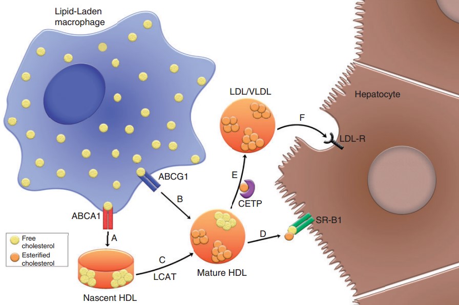 Figure 1. Reverse cholesterol transport
(Source: Allard-Ratick MP, et al. 2021)
HDL reduces inflammation in a variety of cell types including endothelial cells and macrophages. In endothelial cells, HDL inhibits inflammation by decreasing activation of nuclear factor-κB (NF-κB) and 3β-hydroxysteroid-Δ24 reductase, activating the cytoprotective enzyme heme oxygenase-1, and inhibiting activation of inflammatory vesicles. They also reduce monocyte inflammation and attenuate the binding of monocytes to adhesion molecules on the surface of activated endothelial cells.
HDL reduces oxidative stress on LDL and other atherogenic lipoproteins by accepting lipid hydroperoxides and detoxifying them to lipid hydroxides, which are then removed from the circulation by the liver. Small HDL inhibits oxidation more effectively than large HDL. This has the potential to offset some of the effects of diminished cardioprotection brought about by low plasma HDL.
Clinical implications in ASCVD
Low levels of HDL-C are strongly associated with an increased risk of coronary and peripheral artery disease, characterized by atherogenic dyslipidemia consisting of high levels of small, dense LDL particles, elevated triglycerides, and increased insulin resistance. The metabolic syndrome is diagnosed clinically by measuring LDL-C, which is used as a guideline for predicting the risk of atherosclerotic cardiovascular disease (ASCVD). The association between HDL-C and ASCVD varies across ethnicity and gender.
Apolipoprotein A-I is associated with atherosclerosis and is inversely associated with ASCVD risk, however, apoA-I does not improve risk prediction beyond non-HDL-C and HDL-C levels, thus limiting its clinical utility as a prognostic biomarker. Other HDL apolipoproteins may improve risk information but need to be validated in longitudinal cohorts, and their effects should extend beyond HDL-C and other standard risk factors.
HDL function is increasingly being used in observational and interventional line studies. In most studies evaluating in vitro cholesterol efflux from macrophages to apolipoprotein-poor serum, impaired efflux has been shown to be associated with a higher risk of coronary atherosclerotic events and recurrent coronary atherosclerosis. This risk may be present only in coronary atherosclerosis and has little to do with cerebral atherosclerosis and ischemic stroke.
Clinical implications in non-cardiovascular diseases
HDL metabolism is directly related to non-cardiovascular diseases such as cancer and diabetes, and epidemiologic studies have shown that lower levels of HDL-C and apoA-I are associated with an increased risk of lung, liver, colorectal, breast, prostate, and hematologic malignancies, as well as with disease progression and diminished response to treatment. Higher HDL-C and apoA-I levels during treatment also predicted improved response and survival rates.
There is a large body of preclinical data supporting the protective role of HDL in preventing hyperglycemia, and increasing levels of HDL-C and apoA-I therapy can reduce the incidence and progression of diabetes, such as the use of CETP inhibitors and infusion of rHDL. The antioxidant and anti-inflammatory functions of HDL are also reduced in diabetic patients, as is the ability to increase NO production, which can also lead to increased cardiovascular risk in these patients.
In summary, focusing on HDL-C levels as a research priority to accurately elucidate the role of HDL in cardiovascular disease as well as in cancer, infection, diabetes, and other diseases, and to determine the role of inflammation and reverse cholesterol transport in these HDL function-disease relationships is critical to translating HDL into diagnostic or therapeutic targets.
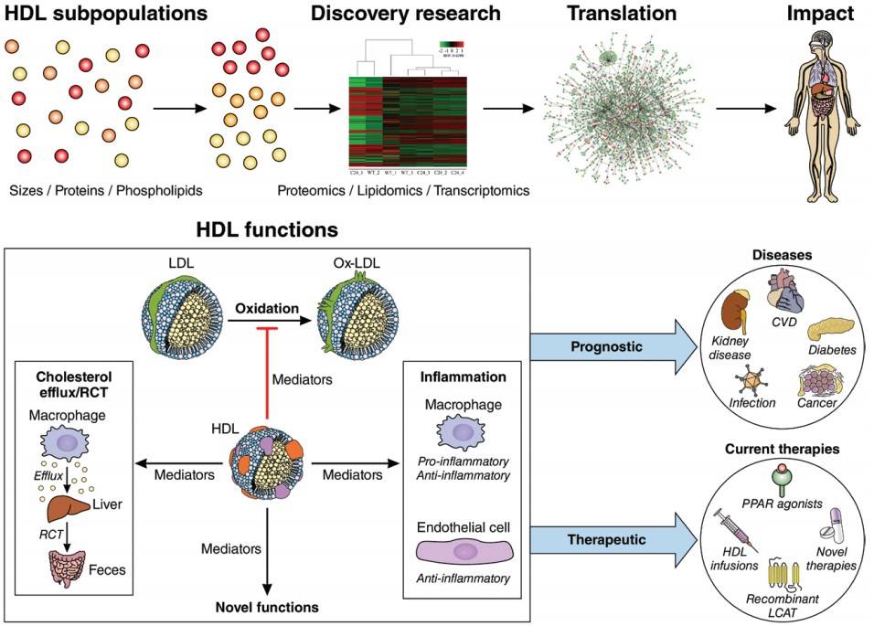 Figure 2. A conceptual framework for investigating the translational and clinical effects of HDLs
(Source: Rohatgi A, et al. 2021)
References
| Target | Cat. No. | Product Name | Size | Species Reactivity | Application | Detection Sample | |
| HDL | DEIA3534 | HDL (Human) ELISA Kit | 96T | Human | Quantitative | Cell culture supernatant, plasma, serum | Inquiry |
| Cholesterol | IKJU-019CL | HDL and LDL/VLDL Cholesterol Assay kit (Colorimetric/Fluorometric) | Quantitative | Serum | Inquiry | ||
| Cholesterol | DEIA-JY2150 | HDL and LDL/VLDL Cholesterol Assay Kit | 192T | N/A | Quantitative | Serum or plasma | Inquiry |
| Target | Cat. No. | Product Name | Expression System | Tag/Conjugate | Application | |
| HDL | DAG-WT2520 | Human HDL control | N/A | Unconjugated | Immunoassays | Inquiry |
| HDL | DAG-WT2522 | High-density lipoprotein cholestero (HDL-C) control | N/A | Unconjugated | Immunoassays | Inquiry |
| Cholesterol | DAG-WT2512 | Total cholesterol (TC) control | N/A | Unconjugated | Immunoassays | Inquiry |
| Cholesterol | DAG-WT2524 | Remnant-like particle cholesterol (RLP-C) control | N/A | Unconjugated | Immunoassays | Inquiry |
| Cholesterol | DAG-WT2525 | Small dense LDL cholesterol (sdLDL-c) control | N/A | Unconjugated | Immunoassays | Inquiry |
| Target | Cat. No. | Product Name | Host | Isotype | Application | |
| HDL | DPABH-16364 | Anti-HDL polyclonal antibody [Biotin] | Rabbit | IgG | WB, IP, RIA, EIA | Inquiry |
| HDL | DPABH-05857 | Magic™ Anti-HDL polyclonal antibody [Biotin] | Chicken | IgY | ELISA, RIA | Inquiry |
| HDL | DMAB6476 | Anti-High Density Lipoprotein monoclonal antibody, clone N-33 | Mouse | IgG2a | N/A | Inquiry |
| Cholesterol | CABT-Z305R | Rabbit Anti-Cholesterol Polyclonal Antibody | Rabbit | IgG | WB, ICC, IHC-P, IHC-F, ELISA | Inquiry |
Loading ......
