
Inquiry Basket
 Loading ......
HSV-1 is a significant pathogen that is ubiquitously carried and transmitted across diverse human populations worldwide. HSV-1's double-stranded genomic DNA can encode over 70 proteins and at least 20 types of miRNAs. In combination with host factors, these molecules can affect HSV-1 lytic, latent, and reactivation infection kinetics.
HSV-1 is an Alphaherpesvirinae with an icosahedral capsid, an envelope and linear double-stranded DNA genome. HSV-1 is known for causing lip, eye and genital infections. HSV-1, a neurotropic pathogen, can also cause herpes simplex encephalitis and neurodegenerative disease.
The envelope is covered by viral glycoproteins. The main glycoproteins responsible for cell binding are glycoprotein B (gB) and gD. Following entry of the HSV-1 genome into the nucleus, viral DNA replication and regulated transcription of the immediate-early (α), early (β), and late (γ) genes takes place. Viral proteins are translated and processed in the cytoplasm; mature structural proteins package the viral genome into new virions, which are transported through the cellular secretory pathways for release, completing a cycle of replication. HSV-1 can cause primary and latent infections, and may be reactivated later. After primary infection by mucosal or epithelial cells, usually with clinical signs and symptoms, HSV-1 establishes latency in sensory neurons in peripheral ganglia. The virus can be reactivated by various environmental stimuli or immune suppression, with resulting clinical signs and symptoms.
 Figure 1. Illustration of the steps involved during HSV entry
(Source: Arii J, et al. 2018)
HSV can enter a broad range of cell types (including neurons, epithelial cells and fibroblasts) in which it expresses its fusion proteins. Following specific binding to receptors on the host cell surface, the viral envelope glycoproteins that mediate entry undergo conformational changes. HSV-1 enters fibroblasts and lymphocytes via direct fusion of the viral envelope with the host plasma membrane. In neurons, however, the virus induces invagination of the plasma membrane to form endocytic vesicles that deliver the virus into the cytoplasm via endocytosis.
Binding of the free virus to the host cell surface is initiated by attachment of the positively charged envelope glycoproteins gC and gB to negatively charged HSPGs, which function to locally concentrate viral particles without eliciting specific membrane fusion. Loss of HSPG binding greatly reduces binding efficiency, but does not block infection, suggesting that this step is not required for entry. The next step in the process is specific recognition and binding of gD to host cell surface receptors, which is the required signal for membrane fusion.
HSV uses three classes of gD receptors: Herpesvirus entry mediator (HVEM), a member of the TNFR family expressed on immune cells, neurons, epithelial cells, and fibroblasts; nectin-1/2, type I transmembrane glycoproteins in the immunoglobulin superfamily—nectin-1, a cell-adhesion molecule used by most alphaherpesviruses across various tissues including neurons; nectin-2, which mediates entry of certain HSV-1 and HSV-2 strains; and 3-O-sulfated heparan sulfate (3-OS HS), a highly sulfated form of HS that serves as an HSV-1 entry receptor.
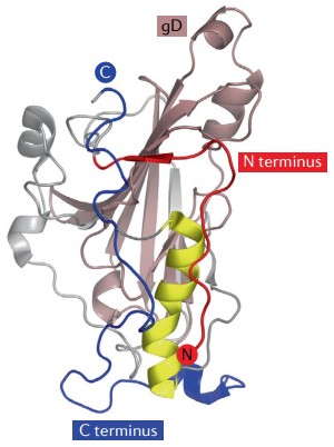 Figure 2. Herpes simplex virus 1 glycoprotein D crystal structures
(Source: Connolly SA, et al. 2021)
gH, encoded by the UL22 gene, is a type I membrane protein of 838 amino acids, with a large extracellular domain, a single transmembrane segment, and a 14–amino-acid cytoplasmic tail. gL, encoded by the UL1 gene, is a 224–amino-acid protein lacking a transmembrane region and cannot anchor to the membrane on its own. Therefore, gH and gL always form a stable 1:1 heterodimer on infected cells and mature virions.
The N-terminal H1 and H2 domains of gH cradle gL, with the H1 domain fitting snugly into a V-shaped groove atop gL. The interacting surfaces of H1 and gL are highly complementary, indicating that each subunit requires the other for proper folding. In the absence of gL, newly synthesized gH is retained in the endoplasmic reticulum and fails to incorporate into the viral envelope. Without gH, some gL is secreted from the cell but cannot anchor to virions, while most remains in an immature form intracellularly. The gH–gL heterodimer's structure differs from known viral fusion proteins, suggesting it does not mediate fusion directly but acts as a fusion regulator. Studies have shown that multiple domains of gH–gL bind distinct neutralizing antibodies, indicating that gH–gL must interact with various viral membrane proteins to transmit the fusion trigger from gD to gB at different sites.
gH–gL acts as an intermediary signaling complex whose interactions with other viral membrane proteins have long been elusive. Studies suggest that the gD–gH–gL binding site may involve the N-terminal region of gH and the C-terminal region of gL, with gH's N-terminus partially shielding gL's C-terminus until gD binds. The cytoplasmic tail of gH (C-terminus) likely interacts with the cytoplasmic tail of gB to regulate and activate the core fusion machinery; insertions or truncations in gH's tail directly impair fusion activity. Thus, one activation pathway may be direct physical association of gH–gL with gD and gB. Additionally, gH–gL binding to its high-affinity integrin receptors αvβ6 and αvβ8 facilitates HSV entry, and engagement with αvβ3 or Toll-like receptor 2 triggers NF-κB signaling and innate immune responses.
gB is a class III fusion protein and the most conserved membrane–fusion glycoprotein in herpesviruses. Anchored in the viral envelope in a prefusion conformation, gB undergoes a major refolding into a postfusion state upon receiving activation signals. The postfusion form exposes hydrophobic residues that insert into the host cell membrane, promoting fusion-pore formation and driving merger of viral and cellular membranes. Studies suggest that the energy for fusion derives from refolding from the high-energy prefusion to the low-energy postfusion conformation.
 Figure 3. Crystal structure of full-length postfusion HSV-1 gB
(Source: Connolly SA, et al. 2021)
X-ray crystallography of the postfusion ectodomain reveals a trimeric, spike- or rod-like structure. Each gB monomer comprises five hairpin-folded domains that interlock with equivalents on adjacent subunits to form a stable trimer. Domain I, the fusion domain, lies near the transmembrane segment and contains two fusion loops. Domain II, the gH–gL binding domain, harbors the binding site for the gH–gL complex. Domain III, made of α-helices, forms the central trimeric coiled coil. Domain IV, or the crown domain, sits at the top of the postfusion trimer and creates a hydrophobic channel that mediates membrane merger. Domain V consists of an extended arm that wedges into grooves between the other protomers, holding the trimer together. The upper portions of domains II and I each include tandem pleckstrin-homology (PH) folds, which may bind lipids.
In addition to activation by gH–gL, gB must bind specific receptors to drive HSV fusion. Paired immunoglobulin-like type 2 receptor α (PILRα) primarily mediates HSV-1 entry into host cells. Expressed mainly on immune cells such as monocytes, macrophages, and dendritic cells, PILRα was the first identified gB receptor. In CHO cells, gB binding to PILRα redirects HSV-1 entry from endocytosis to direct fusion. Other gB receptors include myelin-associated glycoprotein (MAG) on glial cells and nonmuscle myosin heavy chain-IIA (NMHC-IIA), which is expressed predominantly in the cytoplasm.
References
| Target | Cat. No. | Product Name | Size | Species | Application | Detection Sample | |
| HSV | DEIAFY104 | Herpes Simplex Virus Type 1 (HSV-1) Antigen ELISA Kit | 2 plates | Quantitative | Complex sample matrices | Inquiry | |
| DEIA3555 | Mouse/Rat HSV-1 IgG ELISA Kit | 96T | Mouse, Rat | Qualitative | Serum, plasma | Inquiry | |
| DEIA345 | Human Herpes Simplex Virus1 IgA (HSV-1 IgA) ELISA Test kit | 96T | Human | Qualitative | Serum | Inquiry | |
| DEIA1715 | HSV-1 and HSV-2 IgM ELISA Kit | 96T | Human | Qualitative | Serum | Inquiry | |
| DEIA05535 | HSV Type 1 rec. gG1 IgG-ELISA Kit | 96T | Human | Qualitative | Serum, citrate plasma | Inquiry | |
| DEIA344 | Herpes simplex Virus 1 IgG ELISA Kit | 96T | Human | Quantitative | Serum, plasma or cerebrospinal fluid | Inquiry | |
| DEIA346 | Herpes simplex Virus 1 IgM ELISA Kit | 96T | Human | Quantitative | Serum, plasma or cerebrospinal fluid | Inquiry | |
| DEIA350 | Herpes 1/2 IgG ELISA kit | 96T | Human | Quantitative | Serum, plasma or cerebrospinal fluid | Inquiry | |
| DEIA351 | Human Herpes 1/2 IgA ELISA kit | 96T | Human | Quantitative | Serum, plasma or cerebrospinal fluid | Inquiry | |
| DEIA352 | Herpes 1/2 IgM ELISA kit | 96T | Human | Quantitative | Serum, plasma or cerebrospinal fluid | Inquiry | |
| DEIA-NS2401-13 | Herpes simplex Virus 1 and 2 IgG ELISA Kit | 96T | Human | Quantitative | Serum, plasma or cerebrospinal fluid | Inquiry | |
| DEIA-NS2401-14 | Herpes simplex Virus 1 and 2 IgM ELISA Kit | 96T | Human | Quantitative | Serum, plasma or cerebrospinal fluid | Inquiry |
| Target | Cat. No. | Product Name | Expression System | Tag/Conjugate | Application | |
| HSV | DAG-WT2448 | Recombinant HSV-1 Thymidine kinase [His] | E. coli | His | ELISA | Inquiry |
| DAG-T1109 | HSV type 1 (aa 266 - 394) | E. coli | Unconjugated | N/A | Inquiry | |
| DAG-T1112 | HSV type 1 (aa 525 - 578) | E. coli | Unconjugated | N/A | Inquiry | |
| DAG-T1113 | HSV type 1 (aa 84 - 175) | E. coli | Unconjugated | N/A | Inquiry | |
| DAG3048 | Recombinant HSV type 1 | E6 cells | Unconjugated | IA | Inquiry | |
| DAG181 | Native HSV type 1 | N/A | Unconjugated | ELISA | Inquiry | |
| HSV gD | DAG-WT783 | Recombinant HSV-1 Glycoprotein D (a.a. 26-339) [His] | HEK293 cells | His | ELISA, WB, DB | Inquiry |
| DAGC739 | Recombinant HSV-1 Glycoprotein D (a.a. 226-394) [His] | E. coli | His | ELISA, WB | Inquiry | |
| HSV gG | DAG1357 | Recombinant HSV type 1 Glycoprotein [GST] | E. coli | GST | ELISA, WB | Inquiry |
| DAG2013 | Recombinant HSV type 1 Glycoprotein G (a.a. 34-200) [His] | HEK293 cells | His | WB | Inquiry |
Loading ......
