
Inquiry Basket
 Loading ......
Immunofluorescence (IF) is a laboratory technique that allows the detection and localization of a wide variety of antigens in different types of tissues and cell preparations. This method has also been a useful tool for research and clinical diagnostics. IF allows for excellent sensitivity and amplification of signal in comparison to immunohistochemistry, employing various microscopy techniques, such as fluorescence microscopy.
This technique was pioneered by the American pathologist and immunologist, Albert H. Coons (1912–1978) and coworkers in 1942, who demonstrated the localization of antigens in tissues and cells using antibodies against Streptococcus pneumoniae labeled with fluorescein and visualized under ultraviolet light (fluorescence microscopy). By 1950, the development of IF techniques allowed the demonstration of deposition of gammaglobulin in kidney biopsies by Coons and Kaplan as well as complement deposition by Burkholder in 1961. Since then, IF has been incorporated in the diagnostic protocol for assessment of renal biopsy specimens and has become a gold standard technique in the field of renal pathology. For the following 25 years, the Coons's method had been used with different modifications, including the labeling of antibodies with heavy metals, and introducing the use of enzyme-labeled antibodies that lead to the application of this technique in formalin-fixed, paraffin embedded (FFPE) tissues; this method opened the door to the use of immunoperoxidase for routine diagnosis in anatomic pathology.
IF plays an important role in the histopathologic diagnosis of certain diseases affecting eye, skin, heart, brain, and kidney, among other organs. This technique can also be applied in the evaluation of cells in suspension, cultured cells, fresh-frozen tissue, FFPE tissue, beads, and tissue microarrays (TMA) for the detection of specific proteins.
In IF techniques, antibodies are chemically conjugated to fluorescent dyes such as fluorescein isothiocyanate (FITC) or tetramethyl rhodamine isothiocyanate (TRITC). In similar fashion to the enzymatic methods, these labeled antibodies can be used directly (direct IF or primary) or indirectly (indirect IF or secondary) to bind to the antigen of interest, which allows for antigen detection through fluorescence techniques. The degree of fluorescence can be quantified using a flow cytometer, array scanner, automated imaging instrument, or multispectral imaging or visualized using fluorescence or confocal laser scanning microscopy.
There are two methods available, depending on the scope of the experiment or the specific antibodies in use: direct IF (DIF or primary IF) or indirect IF (secondary IF).
DIF uses fluorescent-tagged antibodies to bind directly to the target antigen in the tissue. For example, in autoimmune bullous diseases, the target antigen is the autoantibody or complement that causes the blister. Some of the advantages of DIF includes having a fast and simple methodology process that allows the use of antibodies from the same species. However, it is less flexible and produces a low signal compared to the indirect technique.
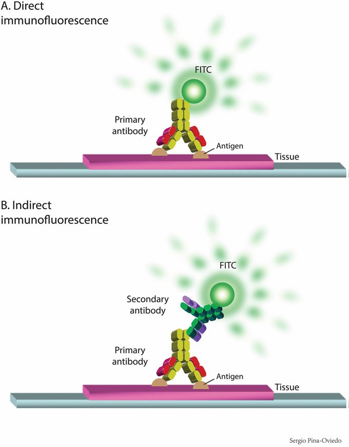 Figure 1. Direct and indirect immunofluorescence methods.
Indirect IF utilizes a two-step technique, in which a primary unlabeled antibody binds to the target, after which a fluorophorelabeled second antibody (directed against the Fc portion of the primary antibody) is used to detect the first antibody. Hence, this is a more flexible technique due to the array of fluorescents used to color secondary antibodies. This technique is more complicated and time consuming than DIF because it requires a second incubation period. However, it is more sensitive because more than one secondary antibody can bind to each primary antibody, which amplifies the fluorescence signal. It is important to notice that the latter can complicate the visualization of the specimen since the background may be also amplified.
Fluorescence and phosphorescence are both types of luminescence. When molecules with luminescent properties absorb light, they emit light of a different wavelength. This emission of light occurs extremely rapid with fluorescence, whereas with phosphorescence the emission continues for milliseconds to minutes after the energy source has been removed.
Fluorescent materials give off light because of their atomic structure. The nucleus of the atom is surrounded by electrons that are arranged in discrete levels of energy, in which each level has a predetermined amount of energy. When an electron absorbs the energy from a photon of light it becomes "excited" and jumps to a higher level of energy that is less stable. This excited state has a half-life of less than 10 s. The electron loses a small amount of energy as heat, and the remainder of the extra energy is given off in the form of a photon. The emitted fluorescence has a lower energy than the absorbed light, so the wavelength of the emitted light is longer than that of the excitation light.
A range of wavelengths of light can excite the electrons of a fluorochrome. For example, fluorescein will fluoresce when hit by light with any wavelength between 450 nm and 520 nm. However, the closer the excitation wavelength is to 495 nm, the more fluorescence will be produced. This optimal wavelength is called the "excitation peak." Similarly, the light produced by fluorochromes has a range of wavelengths that varies from 490 nm to 630 nm, and the emission peak is approximately 515 nm. This phenomenon of fluorescence was first explained in 1852 by Sir George Stokes (1819–1903), a British physicist and mathematician. Hence, the shift in wavelength from short to long during fluorescence is called the "Stokes shift". Some fluorochromes have a small Stokes shift, while others have large Stokes shift. For instance, the fluorochrome fluorescein can be excited by blue-green light, and its Stokes shift is only about 20 nm, which means that the light emitted is color green. On the other hand, the fluorochrome phycoerythrin (PE) can also be excited by blue-green light, but it has a large Stokes shift and the light emitted will be yellow.
One of the most significant problems that most fluorescence-based techniques encounter is photobleaching. This phenomenon occurs when the fluorophore losses its ability to fluoresce due to the generation of reactive oxygen species in the specimen as a byproduct of fluorescence excitation. Photobleaching can be minimized by decreasing the excitation light in both intensity and duration, reducing the availability of singlet oxygen (1O2) by the addition of singlet oxygen scavengers (i.e., antifade reagents), and using a low concentration of a fluorochrome with high-quantum efficiency. For this reason, it is important to keep samples in the dark when not in use, use only as much light as necessary from the microscope, and use an antifade agent in the mounting medium, such a p-phenylenediamine.
Autofluorescence is an intrinsic fluorescence generated directly by the tissue itself and is far brighter in formalin-fixed tissues than in frozen tissues. Hence, clinical kidney IF is performed on frozen rather than FFPE tissues. One of the properties of tissue autofluorescence is that it is present regardless of the wavelength of excitation, although ultraviolet excitation and blue excitation cause more autofluorescence than red excitation. Because the autofluorescence emission is extremely broad regardless of excitation wavelength, it is virtually impossible to find one emission filter for a given excitation wavelength for which there is no autofluorescence present.
The detection of fluorescence probes in tissues and cells sometimes can be problematic due to the presence of flavin coenzymes (FAD and FMN: absorption, 450 nm; emission, 515 nm) and reduced pyridine nucleotides (NADH: absorption, 340 nm; emission, 460 nm) that can produce biological autofluorescence in mammalian cells. Similarly, certain types of transport media or fixatives such as aldehydes -particularly glutaraldehydecan result in high levels of autofluorescence. This situation can be minimized in fixed cells by washing with 0.1% sodium borohydride in phosphate-buffered saline prior to antibody incubation.
Autofluorescence can also be minimized by selecting probes and optical filters that maximize the fluorescence signal relative to the autofluorescence. Other factors that limit IF include the performance of the detection instrument (i.e., setting and calibration of the microscope), the specificity of the antibodies, and the specimen preparation. The recognition of autofluorescence phenomena is necessary to avoid misinterpretation in a biopsy. For instance, masking of the specific fluorescence by the naturally occurring fluorescence of tissues can be troublesome in those type of specimens rich in elastic tissue. However, elastic fibers can be helpful in the recognition of those cases with underlying arterial disease. The background staining can be challenging in certain type of tissue leading to misinterpretation of certain diseases. For instance, a kidney biopsy from a diabetic patient may elicit a linear accentuation of IgG along the glomerular basement membranes, which can be mistaken as an anti-glomerular basement membrane disease. Last, IF on formalin-fixed tissue after enzyme digestion may lead to false positive results if autofluorescence due to staining artifact is not recognized. For example, this artifact can occur in renal biopsies in which the fluorescence can be limited to the capillary lumina and visceral epithelial cells (podocytes) with any anti-sera used.
When measuring fluorescence of more than one color there might be the possibility of overlapping emission signals. This overlapping effect needs to be removed to avoid false levels of signals (overestimation of the actual signal) for one or more colors. This process is called fluorescence compensation and can be automatically calculated in many systems using single positive controls.
Applications of IF in Pathology
1. Sections are thawed at room temperature and dried for 30 min.
2. Immerse the slides in wash buffer at room temperature for 5 min.
3. Remove the excess of wash buffer.
4. Add 100–400μL diluted antibody (e.g., fluorescein-labeled goat anti-human immunoglobulins) and incubate in a moist chamber for 20–30 min at room temperature.
5. Remove the excess of anti-serum by using wash buffer: Immerse the slides in wash buffer two times for 5 min at room temperature.
6. Mount with fluorescence mounting medium and cover with coverslip.
7. Examine slides under a dark-field ultraviolet (IF) microscope.
1. Sections are thawed at room temperature and dried for 30 min.
2. Immerse the slides in wash buffer for 5 min at room temperature.
3. Remove the excess of wash buffer.
4. Add 100–400 μL diluted primary antibody (e.g., anti-human C4d, anti-human C5-9) and incubate in a moist chamber for 20–30 min at room temperature.
5. Remove the excess of antiserum by using wash buffer: Immerse the slides in wash buffer two times for 5 min each at room temperature.
6. Add 100–400 μL of diluted secondary antibody (e.g., goat anti-mouse IgG (H+L)-Alexa Fluor 488) and incubate in a moist chamber for 20–30 min at room temperature.
7. Remove the excess of anti-serum by using wash buffer: Immerse the slides in wash buffer two times for 5 min each at room temperature.
8. Mount with fluorescence mounting medium and coverslip.
9. Examine slides under a dark-field ultraviolet (IF) microscope.
This immunohistochemical technique is considered the gold standard for evaluation of immune deposits in tissue, especially in medical renal diseases. At the time of biopsy or obtention of tissue, the tissue can be transported to the laboratory in fresh salinesoaked gauze or in Michel's fixative solution also known as Zeus medium. The tissue preparation should be performed as follows:
1. Samples transported in Michel's media are washed in buffer.
2. The tissue is embedded in OCT compound in a suitable tissue mold.
3. Immediately freeze the tissue with liquid nitrogen or isopentane.
4. Cut 2–5 μm serial sections in cryostat and place sections on gelatin-coated slides. If necessary, adjust the temperature of the cutting chamber ±5°C, according to the tissue under study.
5. Follow steps for direct or indirect IF, previously described.
6. Examine slides under a dark-field ultraviolet (IF) microscope.
When frozen tissue is inadequate (e.g., lack of glomeruli in kidney biopsies) or not available, immunofluorescence can be performed on FFPE tissue (IF-P) after antigen retrieval. Since the 1970s this technique has become a valuable salvage technique, especially in renal pathology. Based on recent data, IF-P is more sensitive than IF-F for certain kidney lesions and can be used as an unmasking technique. This technique has been proven to be helpful in membranous-like glomerulopathy with masked IgG kappa deposits and membranoproliferative glomerulonephritis with masked monotypic immunoglobulin deposits. The masked effect seen on FFPE tissue is due to the protein cross-linking that blocks antigenicity. Therefore, an antigenretrieval step, which can be performed by either adding a proteolytic enzyme or by heat, is necessary to allow an increased penetration of antibodies to the antigens that are "masked" by formalin fixation.
IF-P is also useful on suspected cases of crystalline light chain proximal tubulopathy and crystalglobulin-induced nephropathy associated to crystalglobulinemia or cryocrystalglobulinemia. In addition, the latter syndromes affecting other organs besides kidney can be demonstrated by the monoclonal staining of crystals; for example, in corneal biopsy samples in which frozen tissue is usually not available, IF-P may help to establish the diagnosis of paraproteinemic keratopathy.
Some of the limitations and pitfalls of this technique have been describe in several studies from kidney biopsies where the IF-P has lower sensitivity than IF-F for detection of C3 deposits, primary membranous nephropathy and antiglomerular basement membrane (GBM) nephritis. Additionally, FFPE tissue might have problems of autofluorescence.
Multiple proteolytic enzymes have been used for antigenretrieval in IF-P including trypsin, Pronase E (protease XIV), proteinase XXIV, and proteinase K. The two most common antigen-retrieval techniques by proteolytic enzymes are the Pronase digestion which was originally described by Fogazzi et al. in 1989, and the proteinase K technique. Heat-induced antigen retrieval, also known as heat-induced epitope retrieval (HIER), can be performed with pH 6.1 citrate or Tris buffers as well as with dual microwave heating using pH 9.0 EDTA antigen-retrieval solution. This step is followed by a cooling-off period and, subsequently, the slides are incubated with FITC-conjugated antibodies against the antigens.
IF controls should include a negative control which consists in a section that does not contain any added fluorophore (omitting primary antibodies): a negative autofluorescence control. And, it should also include a positive control (e.g., albumin) for each run.
Internal controls exist for many of the reagents in routine use. For example, in kidney biopsies, IgA is generally present in tubular casts, IgG in tubular protein resorption droplets and C3 in blood vessels.
Reference
Loading ......
