Introduction of ER+ Breast Cancer Mammosphere Formation and Analysis
In breast cancer, small populations of cells termed breast cancer stem cells (BCSC) have been identified, and in recent years, the concept that these cells are responsible for tumor formation and growth has gained considerable traction. BCSCs have cell surface markers that allow for their identification and recent studies have correlated these markers with prognostic value in triple negative breast cancer. Correlations in other subtypes of breast cancer are less well established and the generalized landscape of BCSC heterogeneity is poorly understood.
Mammosphere formation assays are the foundation to understanding the contribution of BCSC to tumor formation and provide a tractable model system in which to study BCSC proliferation, the contribution of pathways to BCSC activity, and the role of BCSC in the tumor microenvironment. The formation rate of mammospheres correlates well to tumorigenicity of a parental tissue when compared to limiting dilution studies in mouse xenografts and because of this, the number of mammosphereforming cells in a population is thought to directly reflect the numbers of BCSC. Mammospheres form in varying sizes from 50 to 300 μm and the size is thought to reflect proliferative activity of BCSC in a population. Mammosphere formation assays are used to calculate mammosphere forming efficiency (%MFE) of mammospheres derived from both primary tissue and breast cancer cell lines.
This chapter will detail protocols for generalized mammosphere formation assays from breast cancer cell lines, mammosphere formation from cell lines in estrogen-free conditions, and mammosphere formation in matrices.
The most straightforward method for generating mammospheres is by using the reagents provided by several companies. These are optimized for human mammosphere formation using proprietary formulations. The volumes and protocol below are for mammospheres that will be treated therapeutically with drugs (such as in a low throughput screen), but can easily be adapted to accommodate other treatment modalities such as conditioned medium or antibody-based therapies.
When studying breast cancers and resistance to endocrine therapy, it is often necessary to understand the impact of BCSC in the absence of estrogen and phytoestrogens. Phenol red is a phytoestrogen and many commercial products contain unspecified amounts of estrogens. If performing many large experiments, commercial groups can often produce larger quantities of estrogen-free media. We present an estrogen-free option that is a slightly modified version of the protocol first published by the Clarke group.
Methods of ER+ Breast Cancer Mammosphere Formation and Analysis
Methods should be performed in a tissue culture hood using aseptic technique.
Primary Mammospheres
- Prewarm complete Mammocult (containing estrogen) or complete PRF DMEM/F12 (estrogen free) media in a 37°C water bath.
- Record passage number of cells in notebook. Aspirate culture medium from a 10-cm dish of monolayer breast cancer cells, 70–80% confluent.
- Add 3 ml sterile PBS, swirl to rinse, aspirate PBS.
- Using a sterile cell scraper, scrape cells gently from the dish. Do not trypsinize cells at this stage as this could reduce mammosphere forming efficiency.
- Resuspend scraped cells in 3 ml complete media and transfer to a 15 ml conical tube.
- Centrifuge cells at 300Xg for 3 min at room temperature. Carefully aspirate off the supernatant.
- Resuspend the cell pellet in 1 ml complete media and pipet up/down until you have a single cell suspension.
- Count cells by standard methods or by removing 500 μl cell suspension into a 1.5 ml Eppendorf tube. In a second Eppendorf tube, add 400 μl trypan blue. Add 100 μl cells from the first Eppendorf tube to the tube with trypan blue. Mix well at all steps. Put 10 μl trypan blue + cells into each chamber of hemocytometer and count 8 quadrants.
- Plate 20,000 cells/well in an ultra-low adhesion 6-well plate or poly-HEMA-coated plate (estrogen-free only) in 2 ml volume as shown in the example calculation above. Incubate cultures undisturbed in a 5% CO2, humidified incubator at 37°C for 7 days.
Secondary Mammospheres
- Prewarm complete Mammocult (estrogen containing) or complete PRF DMEM/F12 (estrogen-free), trypsin, and thaw any drug treatments.
- If desired, count the number of primary mammospheres, then calculate the percentage of primary MFE by dividing the number of primary mammospheres (>50 μm in diameter) formed by the number of cells plated and express as a percentage.
- Collect the primary mammospheres from all 6 wells into a single 50 ml conical tube and centrifuge at 300Xg for 3 min.
- Aspirate off as much supernatant as possible without disturbing the pellet. Use caution, the pellet may be very loose. Add 3 ml 1XPBS, centrifuge again 300Xg for 3 min, aspirate off PBS.
- Add 1 ml trypsin to cell pellet and pipet up/down for 2–5 min to get a single cell suspension (this step is critical!). Add 1 ml complete media and mix gently.
- Count cells and plate 10,000 cells/well in an ultralow adherent 6-well plate in 2 ml volume (see Note 9).
- Add drug treatment to wells as required for experiment.
- Incubate cultures in a 5% CO2, humidified incubator at 37°C for 3 days.
- After the 3 days, add a second drug treatment to wells as required for experiment (see Note 10). Incubate cultures in a 5% CO2, humidified incubator at 37°C for 4 days then proceed for mammosphere quantitation.
Mammospheres in Matrices
Often cell lines or tissues contain an abundance of BCSC or cells that aggregate together throughout the duration of the mammosphere forming assay, causing large concatemers instead of discrete mammospheres. In these cases, mammosphere formation cannot be accomplished simply by reducing the number of cells plated or the length of time for the assay. Matrices are often required to maintain separation between cells to get appropriate mammosphere formation.
- Set up primary mammospheres, incubate 7 days in a 5% CO2, humidified incubator at 37°C.
- Prewarm Complete Mammocult or Complete PRF DMEM/ F12, the corresponding media + methylcellulose, and trypsin. Thaw any drug treatments.
- Collect the primary mammosphere culture from all 6 wells into a 50 ml conical tube and centrifuge at 300Xg for 3 min.
- Aspirate off as much supernatant as possible without disturbing the pellet. Note: The pellet may be very loose. Add 3 ml PBS, centrifuge again 300Xg for 3 min, aspirate off PBS.
- Add 1 ml trypsin to cell pellet and pipet up/down for 2 min to get a single cell suspension (this step is critical!!). Add 1 ml Complete Mammocult or Complete PRF DMEM/F12 media and mix.
- Count cells. In a 15 ml conical tube, place 10,000 cells, 2 ml of Mammocult + Methylcellulose (estrogen containing) or PRF DMEM/F12 + Methylcellulose (estrogen free), and any drug treatment (see Note 10). Mix and pipet into a well of a low adherent 6-well dish. Repeat for each well in the experiment. If media + methylcellulose becomes too thick, rewarm at 37°C.
- Incubate cultures in a 5% CO2, humidified incubator at 37°C for 7 days.
- Additional drug treatments may be performed by overlaying drug treatment on top of the methylcellulose. This approach has varying levels of success depending on the properties of compound being used and its ability to penetrate the methylcellulose matrix.
- Image and count mammospheres.
Imaging and Counting: Manual Method
- Generate mammospheres.
- Use 4Xobjective to take photographs on a standard inverted bright field microscope.
- After photographing, draw a grid on the bottom of the 6 well dish and manually count mammospheres >50 μm (most photography software has a measurement tool to evaluate size of mammospheres). Mammospheres may also be collected at this point for further molecular analyses.
- If desired, calculate self-renewal by dividing the number of secondary mammospheres formed by the number of primary mammospheres formed.
- It is important to note both the number and size of the resulting mammospheres.
Imaging and Counting: Cellometer Auto T4
An alternative to manual counting with microscopy is the use of automated systems, although most conventional cell counters do not have the capability to accurately count large mammospheres. The Cellometer AutoT4 is an automated cell counter that utilizes one white light emitting diode (LED) in a transmission microscopy setup to acquire and analyze bright field images from cells contained in a disposable counting slide. As currently marketed, the AutoT4 uses an objective at 4Xmagnification, but can be modified to a 2.5Xobjective for imaging a larger area specifically for mammospheres. Also unique to the Cellometer is the choice of the geometry of the disposable counting slide. The slides typically used for direct cell counting of mammalian cell culture have a height of 100 μm, but deeper slides that have a height of approximately 300 μm are available for counting larger objects. With the 2.5Xobjective and the deeper slides, the Cellometer AutoT4 can be used to image, count, and record sizes of mammospheres from 50 to 300 μm.
- Collect mammospheres from one well of a 6-well dish into a 15 ml conical tube.
- Rinse the well with 1 ml PBS to ensure collection of all mammospheres and add to 15 ml conical tube.
- Centrifuge at 300Xg for 3 min and carefully aspirate supernatant.
- Resuspend mammospheres in 50 μl PBS.
- Pipet the 50 μl mammosphere samples into one of the two chambers on the cell counting slide and allow mammospheres to settle.
- Count. The AutoT4 software then automatically captures 2 bright field images and counts the number of mammospheres to generate concentration, diameter, and size distribution results. The results can be directly saved into Excel for downstream calculations. Images are automatically saved for presentation and/or repeat analyses, if necessary.
Imaging and Counting: Celigo Image Cytometer
Another example of an automated system is the use of a plate-based high-speed, high-throughput imager such as the Celigo Image Cytometer. It is advantageous to image and analyze whole wells of mammospheres to generate total count and morphological readout. The Celigo Image Cytometer employs one bright-field and four fluorescent imaging channels: blue (EX377/50 nm, EM470/ 22 nm), green (EX 483/32 nm, EM 536/40 nm), red (EX 531/ 40 nm, EM 629/53 nm), and far red (EX 628/40 nm, EM 688/31 nm), to perform high-throughput 2D and 3D cell-based assays. Cells and spheroids in standard microplates (6, 12, 24, 48, 96, 384, and 1536 well) can be autofocused using Image- Based Auto-Focus (IBAF) or Hardware-Based Auto-Focus (HBAF). The Celigo software consists of five major steps START, SCAN, ANALYZE, GATE, and RESULTS, where the users can enter general information, setup scan/analysis parameters, perform scan/analysis, and then view/export images and results. The simplest analysis is to directly capture bright field images and automatically count the number of mammospheres in the whole well, as well as measure the size and morphology for every mammosphere. The software automatically identifies and segments all of the mammospheres in the whole well, and generate counts, long axis diameter, and area results. The results can be directly exported into Excel for downstream calculations. Images are automatically saved for repeat analyses, and exported for presentation.
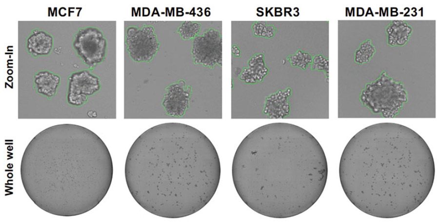 Figure 1. Celigo bright field images of mammospheres.
Immunocytochemistry
Mammospheres can be fixed and immunolabeled as intact spheres to understand how proteins of interest change expression and/or cellular localization following experimental manipulation or drug therapy.
- Set up primary and secondary mammospheres.
- Thaw matrigel overnight at 4°C.
- Prechill Complete Mammocult or Complete PRF DMEM/ F12 and 1.5 ml Eppendorf tubes at 4°C.
- On ice, dilute Matrigel 1:50 with cold media.
- Coat the surface of the chamber slide with diluted matrigel and incubate for 2 h at 37°C.
- Aspirate off any excess Matrigel and return chamber slides to 37°C incubator to solidify Matrigel slide coating.
- Collect mammospheres from the 6-well culture dish and transfer to a conical tube.
- Centrifuge 100Xg for 5 min. Intact mammospheres should be handled very gently in order to avoid dissociation.
- Carefully aspirate the old growth medium and gently resuspend mammospheres in a minimal amount of fresh growth medium. Transfer the mammosphere suspension to a sterile 60- or 100-mm2 dish and select individual mammospheres using a 1 ml pipet (collect as little media as possible).
- Transfer an appropriate number of mammospheres to the Matrigel-coated chamber slides.
- Incubate at 37°C for 1 h to allow mammospheres to attach to the Matrigel.
- Fix by adding room temperature 70% ethanol (**Do not use cold! This will resolubilize the Matrigel!) to the chamber slide and incubating for 15 min at 37°C.
- Aspirate off 70% ethanol and wash one time with room temperature PBS for 20 min. Aspirate PBS then fill chamber slides with fresh PBS. Store at room temperature until immunolabeling, being careful not to let the slides dry out.
Immunolabeling
To generate striking images, mammospheres can be labeled with immunofluorescent secondary antibodies and visualized on a confocal microscope.
- Remove storage PBS from chamber slides, rinse 1Xwith fresh PBS.
- Add primary antibody (if doing dual fluorescence, add both primary antibodies) at recommended dilution and incubate overnight at room temperature.
- Remove primary antibody, rinse with PBS.
- Incubate with fluorescently labeled secondary antibody (both secondary antibodies for dual) at 1:100 for 1 h at room temperature in the DARK.
- Remove secondary antibody, rinse with PBS.
- Mount with a DAPI-containing, aqueous-based mounting media.
- Visualize and capture images using a confocal microscope.

 Loading ......
