Introduction of ELISPOT to Study Stem Cells' Cytokine Secretion
Mesenchymal stem cells (MSCs) are nonhematopoietic stem cells and are capable of differentiation into mesodermal lineages as well as endodermal and neuroectodermal lineages. In addition, recent studies indicate that these cells exhibit immunomodulatory effects on various immune cells, including dendritic cells (DCs), macrophages, and B and T cells, both in vitro and in vivo. The study of the immunosuppressive effect of MSCs on T cells may yield a potential cell therapy for diseases caused by aberrant T-cell activation, such as acute graft-versus-host disease (GVHD). Several studies have demonstrated that the immunosuppressive effects of MSCs are mediated by either direct cell-to-cell contact or by soluble factors. The latter are either secreted from MSCs or upregulated in the immune cells by the presence of MSCs; the MSC-associated factors include interleukin-10 (IL-10), transforming growth factor-B 1 (TGF-B 1), hepatocyte growth factor (HGF), prostaglandin E2 (PGE2), nitric oxide (NO), and indoleamine 2,3-dioxygenase (IDO). However, the mechanism by which MSCs mediate immunosuppression using soluble factors remains controversial. We report that the presence of conditioned media (CM) derived from MSCs in a mixed lymphocyte reaction (MLR) increased the number of IL-10-secreting splenocytes, as measured by an ELISPOT assay, and that the use of neutralizing antibodies to block the IL-10 signaling partially abrogated the immunosuppressive effect of MSC CM.
Methods of ELISPOT to Study Stem Cells' Cytokine Secretion
Immunosuppressive effects of MSCs were mediated by either cell to-cell direct interaction or indirectly by soluble factors released from MSCs or effector immune cells. In an earlier study, we found that the immunosuppression activity of MSCs was largely mediated by soluble factors; therefore, we simplified the assay system by using CM obtained from actively growing MSCs. Other studies reported that the production of suppressive cytokines, such as TGF-B 1 or IL-10, was promoted by MSCs. We understand that the main mechanism underlying this cytokine-mediated immunosuppression involves the downregulation of many cytokines required for T-cell proliferation or survival. We found that the ELISPOT assay is well-suited for finding potential suppressive mediators as well as measuring changes in a full spectrum of cytokines produced during T-cell activation (in an MLR) in the presence of MSC CM.
It is important to note that an independent ELISA should be done to confirm the increase in cytokine concentration and exclude the possibility of contributing effects of the MSC CM itself. We used an ELISA assay to confirm increased secretion of IL-10. Most successful results in ELISA and ELISPOT assays depend on good pairs of antibodies recognizing different epitopes: one for capturing target cytokines in the sample with high affinity and the other for detecting the cytokines. Many good antibody pairs for mouse and human cytokines and chemokines are already commercially available.
Preparation of CM from MSCs and Fibroblasts
BALB/c MSCs or fibroblasts (2 × 10⁶ cells from each) are seeded in a 100-mm culture dish and are cultured for 72 h in complete media. When the plate reaches about 90% confluence, culture supernatant is harvested and centrifuged at 1,200 × g at room temperature for 10 min. Ninety percent of the upper aqueous portion is carefully withdrawn, aliquoted in 1.5-mL microtubes, and kept at −70°C (see Notes 1 and 2).
Preparation of Splenocytes
- 8–12-Week-old BALB/c or C57BL/6 mice are used. After sacrifice by cervical dislocation, fur on the left side is sterilized using 70% ethanol and a small incision is made along the left side of the animal.
- The body cavity is cut open and the spleen is removed using forceps and placed into 10 mL of cold DMEM containing 50 μg/mL gentamicin in a 100-mm culture dish.
- Spleen is cut into 5–6 pieces using scissors.
- Pieces of spleen are mechanically homogenized between the frosted slides.
- Homogenized spleen is passed through a cell strainer mounted on a 50-mL conical tube.
- Cell strainer is washed with 5 mL of complete media.
- The conical tube is spun at 800 × g for 10 min.
- The supernatant is discarded, and the pellet is resuspended in 1 mL of red blood cell lysis buffer and incubated at room temperature for 5–10 min.
- Roughly, 9 mL of complete media is added to the conical tube and spun as in step 7.
- The supernatant is discarded and the pellet is resuspended in 3 mL of complete media.
- 10 μL of the cell suspension is diluted with trypan blue and counted using a hemocytometer to determine the total splenocyte cell count (see Note 3).
ELISPOT
- The day prior to the ELISPOT experiment, each well of a 96-well ELISPOT plate is soaked with 15 μL/well of 35% ethanol for 1 min and then washed twice with 200 μL of PBS per well before ethanol evaporation (see Note 4).
- The plate is coated with 100 μL of capture anti-IL-10 monoclonal antibody per well (the antibody is diluted 1:60, antibody to PBS, in sterile PBS) and incubated overnight at 4°C. The plate is sealed to prevent evaporation during the incubation period.
- On the day of the experiment, the plate is washed once with 200 μL of PBST per well and blocked with 200 μL of complete media per well for 30 min at 37°C.
- BALB/c splenocytes and G-irradiated C57BL/6 splenocytes (20 Gy) are used as responder and stimulator, respectively. Both cell types (5 × 10⁵ cells of each in 100 μL complete media) are cultured with 100 ML of complete media (control), BALB/c MSC CM (MSC CM), or fibroblast CM (Fibro. CM) for 72 h. The controls are as follows: irradiated C57BL/6 splenocytes only, BALB/c splenocytes only, and blank wells.
- After 72 h, the cells are discarded and 200 μL of distilled water is added to each well. The plate is then placed on ice for 10–15 min (see Notes 5 and 6).
- The plate is washed five times with 200 μL of PBST per well.
- 100 μL of detection antibody, diluted 1:60 in PBS, is added to each well; the plate is then incubated for 1 h at 37°C (see Note 7).
- The plate is washed five times with 200 μL of PBST per well, and 100 μL of streptavidin–alkaline phosphatase, diluted 1:2,000 in PBST–1% BSA, is added to each well and incubated for 1 h at 37°C.
- The plate is washed four times with 200 μL of PBST per well and twice with 200 μL of PBS per well (see Note 8).
- 50 μL of developing solution, composed of 20 μL/mL of NBT/BCIP stock solution in an alkaline phosphatase substrate buffer, is added to each well and color development is carefully monitored for 5–30 min.
- When the experiment yields a reasonably high ratio of signal to noise, the plate is washed with tap water and dried by blotting with absorbent paper.
- The resulting spots are counted and analyzed on a computer-assisted AID ELISPOT Reader System.
ELISA
- A 96-well Maxi-sorb plate is coated with 100 μL of capture antibody diluted 1:200, antibody to PBS, in PBS per well and stored overnight at 4°C. The plate is tightly sealed to prevent evaporation during the incubation period.
- On the day of the experiment, the plate is washed five times with 200 ML of PBST per well and incubated with 200 μL of 1× assay diluent per well for 1 h at room temperature.
- The plate is washed five times with 200 μL of PBST per well.
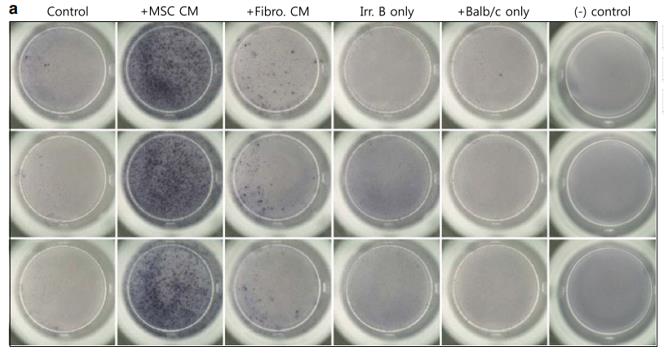 Figure 1. Measurement of IL-10-secreting cells in MLR by ELISPOT assay.
- 100 μL of standard mouse IL-10 is serially diluted in 1× assay diluent and combined with experimental samples from the MLR (cell-free supernatant) or the MLR with MSC CM; the solutions are then added to separate wells on the ELISA plate and incubated for 2 h at room temperature (see Note 9).
- The plate is washed five times with 200 μL of PBST per well.
- 100 μL of detection antibody, diluted 1:60 in PBS, is added to each well; the plate is then incubated for 1 h at room temperature.
- The plate is washed five times with 200 μL of PBST per well.
- 100 μL of avidin–HRP, diluted in 1× assay diluent at 1:2,000, is added to each well and the plate is sealed and incubated for 30 min at room temperature.
- The plate is washed seven times with 200 μL of PBST per well.
- 100 μL of the substrate solution is added to each well and incubated for 15 min at room temperature.
- 50 μL of stop solution is added to each well.
- Absorbance is read at 450 nm with background subtraction at 590 nm.

 Loading ......
