
Inquiry Basket
 Loading ......
Reactive oxygen species (ROS), such as hydrogen peroxide (H2O2), can be generated in live cells and tissues either via normal oxidative intracellular metabolism or induced by extracellular toxins which inhibit activity of antioxidant enzymes. Reduced endogenous antioxidant capacity causes natural overproduction of ROS which, in turn, leads to oxidative stress. Hydrogen peroxide can affect different biochemical reactions in human lymphocytes, including alterations in enzymatic activities, lipid peroxidation, and damage to DNA, and it was reported that short-term exposure of lymphocytes to hydrogen peroxide suppresses NFK B, AP-1, and NFAT transcription factors which play important roles in regulating production of cytokines.
Enzyme-linked immuno spot (ELISPOT) allows detection of just a few cytokine-releasing cells out of tens of thousands making this technique a method of choice for vaccine development, cancer research, AIDS research, allergy research, and autoimmune disease studies. We find that ELISPOT assay can be easily adopted for studying oxidative stress in human lymphocytes and, if combined with multidonor testing, ELISPOT can be used for high-throughput screening of multiple oxidative stress substances.
The objective of this study was to examine the effect of oxidative stress induced by hydrogen peroxide in unstimulated peripheral blood mononuclear cells (PBMCs) on their capacity to secrete IFN-gamma, IL-2, IL-4, IL-5, IL-8, and TNF-alpha. Our oxidative stress model utilized short-term treatment of lymphocytes cultured in vitro with hydrogen peroxide in a concentration that does not impair lymphocyte viability. We employed ELISPOT assays which are more sensitive than ELISA and permit determination of frequency of cytokine-secreting cells.
Our results indicate that hydrogen peroxide-induced oxidative stress significantly inhibits secretion of TH1 cytokines IFN-gamma and IL-2, as well as proinflammatory chemokine IL-8 and inflammatory cytokine TNF-alpha. It appeared that acute oxidative stress induced by hydrogen peroxide did not affect the frequency of cell secreting TH2 cytokines: the regulator of adaptive and humoral immunity IL-4 and B-cell activator IL-5. Interestingly, there was no linear correlation between the number of cultured cells and the number of spots for each cytokine tested. The lack of such a correlation is not completely understood and additional studies are required to address this issue.
1. Ficoll-Paque PLUS.
2. 50 mM phosphate-buffered saline (PBS), pH 7.2.
3. Red blood cell lysing solution: 155 mM NH4Cl, 10 mM NaHCO3, and 0.1 mM EDTA.
4. RPMI complete culture medium: RPMI1640 (1 L) supplemented with 50 mL of heat-inactivated fetal calf serum, 1.19 g HEPES, 2 g of sodium bicarbonate, 3.5 ML of beta-mercaptoethanol, 50 mg Gentamicin Reagent Solution (see Notes 1 and 2).
5. Centrifuge allowing spinning of 50-mL culture tubes at 500 × g.
6. Hemacytometer to count lymphocytes under the microscope.
7. Trypan blue dye.
8. Upright microscope equipped with bright-field illumination and phase-contrast condenser.
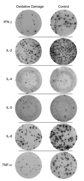 Figure 1. Typical ELISPOT images of secreted cytokines from human PBMCs.
1. Reagent to induce oxidative stress: First, prepare 1 mM solution of H2O2 by adding 0.5 μL of 30% H2O2 to 8.8 mL of Hank's balanced salt solution (HBSS). Then, prepare 10 μM solution of H2O2 by adding 100 μL of 1 mM H2 O2 solution to 9.9 mL of RPMI complete culture medium.
1. Commercially available, ready-to-use ELISPOT assay kits to study secretion of human IFN-G, IL-2, IL-4, IL-5, IL-8, and TNF-α. Each kit includes a dry 96-well, PVDF, membrane backed plate precoated with capture antibody, a concentrated solution of detection antibody, a concentrated solution of streptavidin-conjugated alkaline phosphatase, BCIP/NBT chromogenic substrate, and wash and dilution buffers.
2. Mitogens to stimulate release of cytokines from cultured PBMCs: Calcium ionomycin, Phorbol 12-myristate 13-Acetate, Phaseous Vulgaris Red Kidney Bean Phytohaemagglutinin.
3. Hand-held Nunc-Immuno™ 12-plate washer.
4. Membrane-removal device.
5. ELISPOT plate reader QHub.
1. Collect blood samples from healthy donors in standard citratephosphate-dextrose unit bags and separate PBMCs using density centrifugation (500 × g for 30 min) of 25 mL of blood layered on 20 mL of 1.077 g/mL Ficoll-Paque Plus at 25°C (see Note 3).
2. Discard the upper plasma layer after centrifugation and transfer PBMCs into two sterile 50-mL tubes.
3. PBMCs were then resuspended in 45 mL of sterile PBS and centrifuged for 5 min at 500 × g.
4. Discard supernatant, resuspend the pellet in 10 mL of red blood cells lysing solution, and incubate for 5 min at room temperature.
5. After lysing, add sterile PBS to reach 50 mL graduation mark on the tube to resuspend PBMCs.
6. Centrifuge tubes for 5 min at 500 × g.
7. Discard supernatants and add 30–40 mL of RPMI complete medium to the tubes with PBMCs.
8. Mix cells 1:2 with Trypan blue dye and pipette 10 μL of that mixture into each side of a hemacytometer under a coverslip (see Note 4). Count cells under the microscope using 20× lens and phase-contrast condenser.
1. Add PBMCs directly to RPMI complete culture medium containing hydrogen peroxide and incubate in 37°C/CO2 humidified incubator for 30 min.
2. Transfer PBMCs from the incubator into a sterile hood, discard culture media with hydrogen peroxide, and rinse cells three times with sterile culture media.
3. Prepare several serial dilutions of PBMCs and mix them with corresponding mitogens (see Note 5).
Treatment of human PBMCs with H2O2 caused almost twofold decrease in the number of lymphocytes secreting the TH1 cytokines IFN-gamma and IL-2, as well as chemokines IL-8 and TNF-alpha. However, the number of cells secreting TH2 cytokines IL-4 and IL-5 in hydrogen peroxide-treated group did not change.
Results of this study suggest that oxidative stress can change the balance between TH1 and TH2 cytokine secretion which, in turn, may underlie developments of various pathological conditions. It appears that ELISPOT assay can be used as a convenient research tool for studying the effects of oxidative stress on PBMCs and turned into a high-throughput platform to screen various oxidative stress compounds.
1. Sterilize RPMI complete culture medium and reagents that are used to separate out the white blood cells through 0.2-μm sterile filter to allow their long-term storage.
2. When using fetal calf serum, it is important to heat inactivate the serum at 56° for 30 min. After the heat inactivation, the serum should be filtered.
3. When layering Ficoll, make sure that the blood does not mix with the Ficoll to gain the best separation and highest yield of PBMCs.
4. Overfilling hemacytometer with cell solution may result in inaccurate cell quantification. While counting cells on a hemacytometer, first find the middle square which contains 25 smaller squares and count cells in 5 of them. Calculate the average and multiply by 25 (total number of squares in that area), then multiply by 2 (cell dilution factor), and multiply by 10,000 to determine the number of cells in 1 mL of original cell suspension. The resulting number should be used for calculating serial dilutions of PBMCs.
5. Making serial dilutions of cells allows avoidance of overdevelopment of ELISPOT plate and obtains a quantifiable number of spots that can be counted either manually or using automated ELISPOT plate readers.
6. For better well-to-well reproducibility, cells need to be mixed thoroughly before adding them into the wells. This may require shaking the tube with cells after filling every four wells in ELISPOT plate.
7. Plates can be wrapped into aluminum foil to provide even heat distribution across the bottom of the ELISPOT plates during their incubation. This helps to improve well-to-well spot consistency across the plate. Aluminum foil also helps to reduce a background staining. This is a very simple procedure which can be done as follows: before plating cells, ELISPOT plate is placed onto 13 × 16-cm piece of aluminum foil; after that, cells are added into the wells, plate is covered with the lid, and edges of the foil are shaped loosely around the edges of the plate to wrap it. After finishing the incubation of the cells, the foil can be removed and either discarded or saved and used on the next ELISPOT plate.
8. Shelves in the CO2 incubator must be level to avoid moving cells toward one side of the well: this may produce under- and overdeveloped parts of the well and hinder quantification of spots. It is also important to avoid disturbing cultured cells (e.g., by slamming the door of the incubator) during the incubation which may cause developing of weakly stained fuzzy spots.
9. Make sure that the height of prongs in the handheld plate washer is properly adjusted so that prongs do not touch the membranes on the bottom of the ELISPOT plate: PVDF membranes are fragile and can be easily punctured by protruding prongs.
10. Between washes, it is important to tap out the excess liquid in the well onto a paper towel to prevent diluting the sequential reagents added to the plate.
11. Plates must be completely dried before analysis because wet membranes appear dark and obscure detection and quantification of spots.
Reference
Loading ......
