
Inquiry Basket
 Loading ......
Progesterone is an important hormone that regulates endometrial receptivity, egg maturation, and embryonic development. The endometrium changes under the action of progesterone, providing a suitable environment for embryo implantation. The development, maturation and ovulation of egg cells are inseparable from the role of progesterone. Both in vivo and in vitro embryo development requires the participation of progesterone. There has been controversy about the clinical impact of progesterone, and the relationship between progesterone levels and pregnancy rates cannot yet be fully explained. Progesterone levels vary with fluctuations in its secretion, so analyzing progesterone levels can be difficult. Insufficient levels of progesterone secreted in early pregnancy may be a trigger for miscarriage. The researchers pointed out that although there is not enough evidence to prove that using progesterone in early pregnancy can prevent miscarriage, using progesterone in women with recurrent miscarriage can significantly reduce the miscarriage rate. In assisted reproductive technologies, pregnancy rates using progesterone as a luteal supporter are higher than those in a control group without progesterone. Therefore, in order to prevent miscarriage, embryonic death and premature birth, progesterone drugs have been commonly used in pregnant women, but their pathophysiological mechanisms are still unclear. This article reviews the research progress on the effects of progesterone on the development of endometrium, egg cells and embryos.
 Figure 1. Diagram of the menstrual cycle showing the ovarian (follicular and luteal) and endometrial (proliferative and secretory) phases.(Kalakota NR, et al.; 2022)
The progesterone receptor is a special high-affinity nuclear receptor that can directly regulate the expression of a large number of endometrial genes. It has two subtypes, type A and type B, which are derived from the transcription and translation process of a single gene. Each subtype is capable of direct action. Progesterone exerts its influence on the endometrium through this receptor. In addition, its effect also depends on other signaling factors and transcription factors. Progesterone can cause decidualization of the endometrium. However, the entire decidualization process requires progesterone receptors and cyclic adenosine monophosphate (cAMP) to transmit signals. cAMP induces the expression of many transcription factors, and these transcription factors can directly affects or modulates progesterone receptors. These transcription factors and progesterone receptors together form a multimeric complex that causes gene transcription and translation into a decidualization phenotype. Without the interaction between these cells signaling factors and transcription factors, progesterone would be unable to exert its effects on the endometrial matrix. Progesterone-induced decidualization of the endometrium may serve as a biosensor between the embryo and the uterus during early embryo implantation.
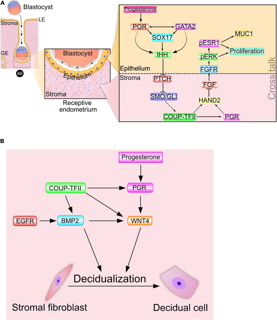 Figure 2. Signaling cross talk drives progesterone-dependent endometrial receptivity and decidualization.(Maurya VK, et al.; 2021)
Progesterone regulates the cyclic proliferation and decidualization of the endometrium during the menstrual cycle, improves the receptivity of the endometrium, and facilitates embryo implantation. Appropriate progesterone levels after fertilization not only promote embryo implantation, but also maintain pregnancy by promoting endometrial development and inhibiting myometrial contraction. However, the time window for the emergence of endometrium that allows the embryo to implant is very short, and this window period is affected by progesterone. The expression and local distribution of progesterone receptors and their co-regulators are different at different stages of the menstrual period. During the implantation window, immunohistochemical staining of progesterone receptors in endometrial epithelial tissue gradually fades. In the middle and late stages of endometrial proliferation, progesterone receptors A and B are easily detected in epithelial tissue and stroma. Progesterone receptor A is not expressed in the epithelial tissue during the secretory phase, while progesterone receptor B is maintained at a low dose and even drops even lower in the late secretory phase. However, in the endometrial stroma, there are always more progesterone receptor A than progesterone receptor B.
During the endometrial secretory phase, progesterone enhances endometrial receptivity and facilitates implantation. However, only a very small amount of progesterone is required during this process. Therefore, during the luteal phase, women with normal endometrial function only need a small amount of progesterone to exert reproductive effects. For those women with infertility caused by endometrium factors, there may be progesterone resistance, and treatment effects can be achieved by increasing progesterone levels, but this hypothesis needs to be confirmed. When studying endometrial gene expression in young healthy women, it was found that high progesterone levels can overexpress many biological processes controlled by genes, such as cell adhesion, immune system and organ development, etc., which can affect the endometrium and embryonic implantation bed. The gene expression in the endometrium after ovulation induction at the end of the follicular phase is different between women with elevated progesterone levels (>4.77 nmol/L) and women with normal progesterone levels, further proving that human chorionic gonadotropin (hCG) High levels of progesterone on the day of injection lead to poorer outcomes in vitro fertilization (IVF) cycles mainly because they affect endometrial receptivity and result in lower implantation rates. Therefore, it is of high value to closely monitor changes in serum progesterone levels during ovulation induction in IVF cycles. When progesterone increases, you can choose to freeze the embryos first and then perform embryo transfer in the natural cycle.
There is currently uncertainty about the role of progesterone on human egg cell maturation and quality. It is known that in the follicular fluid before ovulation, estrogen dominance will transform into progesterone dominance. This process is synchronized with the resumption of meiosis and the maturation of egg cells. It is speculated that progesterone may play a certain role in this process. Steroid hormones, especially progesterone, can cause the egg cell to resume meiosis and further divide to the MⅡ stage. If the effect of progesterone is inhibited, meiosis cannot be restored and the egg cell cannot develop to the MⅡ stage. Administration of progesterone antagonists or its receptor antagonists in experimental mice prevented both luteinizing hormone (LH)-triggered blastocyst rupture and spontaneous maturation of cumulus and egg cell complexes. The content of progesterone in follicular fluid and the ratio to estrogen directly affect the quality and maturation of oocytes. However, the effect of progesterone in the in vitro culture and maturation (IVM) of immature oocytes is controversial. Studies have found that adding different doses of progesterone to mouse GV phase oocytes during IVM cannot improve the maturity rate and developmental ability. When the dose of progesterone is increased from 10 μmol to 100 μmol, the maturity rate is significantly reduced. The rate of developmental arrest in the GV stage increased significantly. The effect of progesterone on inhibiting meiotic resumption was more effective in cumulus oocytes than in cumulus-less oocytes. This may mean that the egg cell has a close interaction with its surrounding cumulus cells, and that the egg cell can affect the function of the cumulus cells.
The low apoptosis rate of cumulus cells is related to the developmental ability of egg cells. During IVM of cumulus oocytes, the lower the apoptosis rate of cumulus cells, the stronger the developmental ability of egg cells. Progesterone acts as a pro-survival factor and anti-apoptotic intermediate mediator in this process.
Progesterone can affect egg cell quality by affecting the development of dominant follicles. During the estrous cycle, the progesterone content in the serum can regulate the pulsatile release of gonadotropin-releasing hormone, which in turn can regulate the pulse frequency of LH secretion. The pulse frequency of LH secretion determines whether a dominant follicle can ovulate. Therefore, when the progesterone content is high, the LH pulse frequency will be very low, resulting in atresia of the dominant follicle. During super-ovulation induction, high-concentration progesterone regulates the pulse frequency of LH, preventing premature maturation of the egg nucleus, thereby improving egg cell quality. In a normal menstrual cycle, the LH pulse frequency increases as the progesterone content decreases after follicle luteinization, stimulating the dominant follicle to continue growing. At the same time, the dominant follicle secretes more estrogen and inhibin, ultimately achieving the purpose of ovulation. Low concentrations of progesterone (3.18-6.36 nmol/L) can increase the LH pulse frequency, but the increase is not enough to reach the pulse frequency that can mature follicles and ovulate, which will lead to a prolonged duration of dominant follicles. As the duration of dominant follicles increases to 4 to 8 days, egg cell quality improves, and the pregnancy rate will also increase. However, if it exceeds 10 days, the pregnancy rate will decrease significantly. Therefore, progesterone affects the development of dominant follicles by regulating the frequency of LH pulses, thereby affecting the quality of egg cells.
The development of embryos in microenvironments such as the fallopian tube and uterus is mainly regulated by progesterone. Early pregnancy failure and poor embryonic development are often related to low progesterone levels. The impact of progesterone on embryonic survival and development is divided into two aspects. On the one hand, progesterone directly affects the growth and development of the embryo as a pro-survival factor. On the other hand, progesterone promotes the secretion of related cell activity factors and indirectly affects the growth and development of the embryo. Progesterone receptors are expressed at all stages of embryonic development, allowing progesterone to directly affect embryonic development. Supplementing progesterone during ovulation can increase the pregnancy rate, but delaying the time of supplementation will invalidate the promotion effect. Therefore, the key to affecting embryonic development is the time of progesterone supplementation rather than the content of progesterone. Under the regulation of progesterone, the embryo and endometrium can secrete a granulocyte-macrophage colony-stimulating factor to promote embryonic development. Progesterone can increase the growth factors secreted by endometrial stromal cells.
References
| Target | Cat. No. | Product Name | Size | Species Reactivity | Application | Detection Sample | |
| 17-OH progesterone | DEIABL241 | 17-Hydroxyprogesterone ELISA Kit | 96T | Quantitative | serum, plasma, urine, fecal extracts | Inquiry | |
| DEIA-XY50 | 17 α-Hydroxyprogesterone ELISA kit | 96T | N/A | Quantitative | Saliva | Inquiry | |
| Hydroxyprogesterone | DEIACL46 | CDSimple™ 17-OHP Chemiluminescent ELISA Kit | 96T, 192T | Quantitative | Serum | Inquiry | |
| Medroxyprogesterone | DEIABL-QB17 | Medroxyprogesterone ELISA Kit | 96T | Quantitative | tissue, liver, fish, shrimp | Inquiry | |
| MedroxyProgesterone Acetate | DEIA-XY30 | Medroxyprogesterone Acetate ELISA kit | 96T | Quantitative | tissue (muscle), fat, beverages, feed | Inquiry | |
| Progesterone | DEIA-BJ2963 | Bovine Pg(Progesterone) ELISA Kit | 96T | Bovine | Quantitative | Serum, plasma, tissue homogenates, other biological fluids | Inquiry |
| DEIASL220 | Progesterone ELISA Kit | 96T | Quantitative | milk, cheese, yogurt, ice cream, butter | Inquiry | ||
| DEIA4101 | PG(Progesterone) ELISA Kit | 96T | Universal | Quantitative | Serum, plasma, tissue homogenates and other biological fluids | Inquiry | |
| DEIA4997 | Progesterone ELISA Kit | 96T, 480T | Quantitative | serum, plasma | Inquiry | ||
| DEIA1591 | Progesterone Human ELISA Kit | 96T | Human | Quantitative | serum | Inquiry | |
| DEIA1592 | Progesterone Human ELISA Kit | 96T | Human | Quantitative | serum, plasma | Inquiry | |
| DEIA2248 | Progesterone ELISA Kit | 96T | Quantitative | serum, plasma | Inquiry | ||
| DEIA2233 | 17-α-OH Progesterone ELISA Kit | 96T | Quantitative | serum | Inquiry | ||
| DEIA284 | Progesterone ELISA Kit | 96T | Human | Quantitative or qualitative | Serum and plasma | Inquiry | |
| DEIA-S10023 | Salivary 17 α-hydroxyprogesterone ELISA Kit | 96T | Quantitative | saliva | Inquiry | ||
| DEIABL256 | Progesterone ELISA Kit | 96T | Quantitative | fecal extracts, urine, TCM | Inquiry | ||
| DEIA-XY2274 | PG(Progesterone) ELISA Kit | 96T | Universal | Quantitative | Serum, plasma, tissue homogenates and other biological fluids. | Inquiry | |
| DEIACL45 | CDSimple™ Progesterone Chemiluminescent ELISA Kit | 96T, 192T | Quantitative | Serum | Inquiry |
Loading ......
