
Inquiry Basket
 Loading ......
Immunocytochemistry (ICC) is popular, simple, affordable, and powerful technique providing the possibility of using specific antibodies to identify abundance and cellular location of target molecules (biochemically defined antigens). It is a perfect complement to biochemical methods (assuming uniformity of samples) as it allows the analysis of single cell differences. ICC samples may be cultured cell lines or cells isolated directly from organism. Staining of paraffin embedded or frozen tissue sections (immunohistochemistry—IHC) requires a different protocol. In both— ICC and IHC antibodies conjugated with chromogenic reagents or fluorochromes (immunofluorescence, IF) are often employed for detection of target antigens. Nonfluorescent detection mostly uses enzyme-conjugated antibodies such as, for example, horseradish peroxidase (HRP) that converts a soluble substrate (e.g., 3,3- 0-diaminobenzidine [DAB]) into colored precipitate at the antigen site. However, growing number of fluorescent labels, ease of use and their vast advantages makes them being chosen over chromogenic detection. Fluorochromes can be used to label primary antibodies in direct detection method or attached to secondary antibodies in two-step indirect detection. Direct method is shorter and simple with minimized species cross-reactivity and nonspecific binding. On the other hand, it results in weaker signal and has less flexibility, as commercially available labeled primary antibodies are limited and usually more expensive. In indirect method cells are exposed to unlabeled primary antibody followed by fluorochrome-conjugated secondary antibody. Because of the additional step of secondary antibody incubation, it is longer and more complicated, especially in experiments with multiple antigens detection. However, utilization of secondary antibodies results in great flexibility and sensitivity, as well as lower costs. Precautions should be taken to prevent cross-reactivity and high background. Here we demonstrate the protocol of LN-229 human glioblastoma cells treatment with rapamycin (autophagy inducer) or chloroquine (inhibitor of autophagosomes degradation) and subsequent double labeling of the key autophagy protein—LC3 (microtubule-associated protein 1A/1B-light chain 3) and IRS-1 (insulin receptor substrate 1) in search of colocalization and previously reported IRS-1/LC3 nuclear structures.
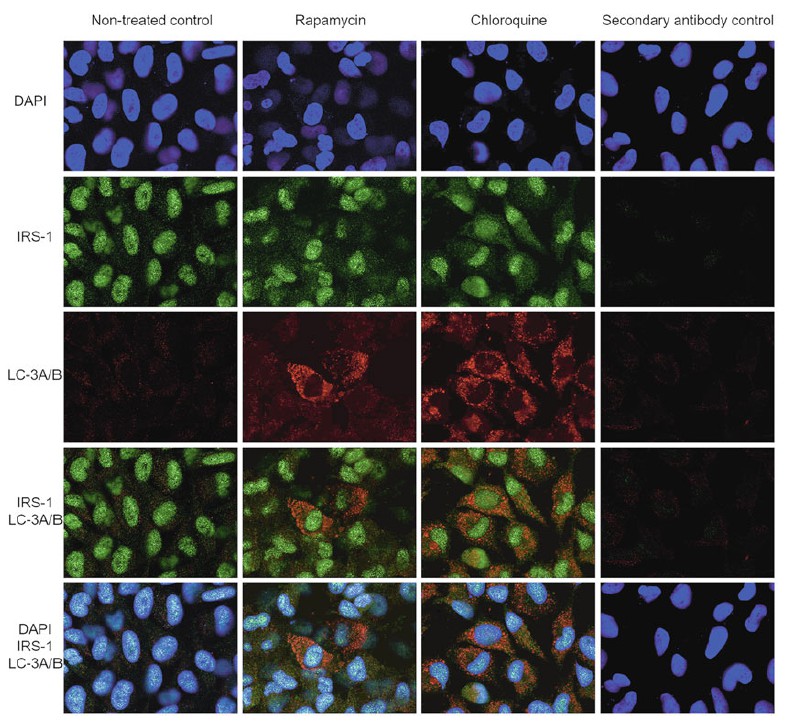 Figure 1. Immunocytochemical detection of IRS-1 and LC3A/B in LN-229 cells untreated and treated with rapamycin or chloroquine for 24 h.
Reference
Loading ......
